7UFJ

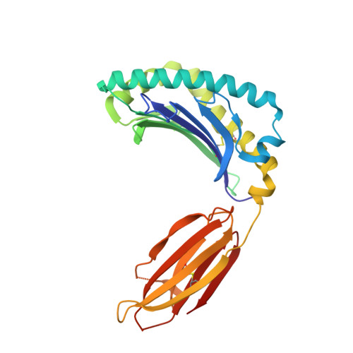
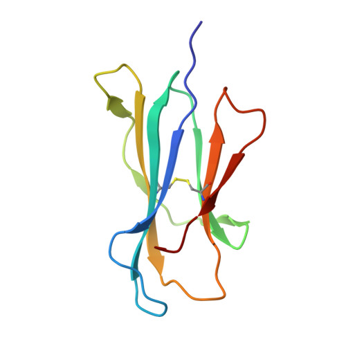
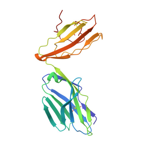
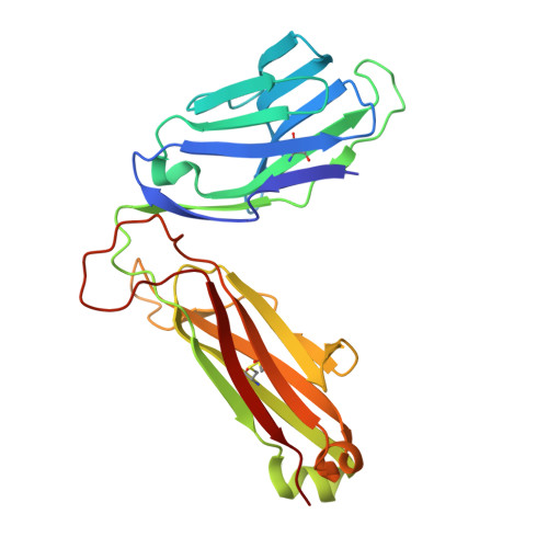
Structure of human MR1-ethylvanillin in complex with human MAIT A-F7 TCR


Experimental Data Snapshot

  • Method: X-RAY DIFFRACTION
  • Resolution: 2.50 Å
  • R-Value Free: 0.238 
  • R-Value Work: 0.180 
  • R-Value Observed: 0.183 

wwPDB Validation   3D Report Full Report


Ligand Structure Quality Assessment 


This is version 1.2 of the entry. See complete history


Literature

Quantitative affinity measurement of small molecule ligand binding to Major Histocompatibility Complex class-I related protein 1 MR1.

Wang, C.J.H.Awad, W.Liu, L.Mak, J.Y.W.Veerapen, N.Illing, P.T.Purcell, A.W.Eckle, S.B.G.McCluskey, J.Besra, G.S.Fairlie, D.P.Rossjohn, J.Le Nours, J.

(2022) J Biol Chem 298: 102714-102714

  • DOI: https://doi.org/10.1016/j.jbc.2022.102714
  • Primary Citation of Related Structures:  
    7UFJ

  • PubMed Abstract: 

    The Major Histocompatibility Complex class I-related protein 1 (MR1) presents small molecule metabolites, drugs, and drug-like molecules that are recognized by MR1-reactive T cells. While we have an understanding of how antigens bind to MR1 and upregulate MR1 cell surface expression, a quantitative, cell-free, assessment of MR1 ligand-binding affinity was lacking. Here, we developed a fluorescence polarization-based assay in which fluorescent MR1 ligand was loaded into MR1 protein in vitro and competitively displaced by candidate ligands over a range of concentrations. Using this assay, ligand affinity for MR1 could be differentiated as strong (IC 50  < 1 μM), moderate (1 μM < IC 50  < 100 μM), and weak (IC 50 > 100 μM). We demonstrated a clear correlation between ligand-binding affinity for MR1, the presence of a covalent bond between MR1 and ligand, and the number of salt bridge and hydrogen bonds formed between MR1 and ligand. Using this newly developed fluorescence polarization-based assay to screen for candidate ligands, we identified the dietary molecules vanillin and ethylvanillin as weak bona fide MR1 ligands. Both upregulated MR1 on the surface of C1R.MR1 cells and the crystal structure of a MAIT cell T cell receptor-MR1-ethylvanillin complex revealed that ethylvanillin formed a Schiff base with K43 of MR1 and was buried within the A'-pocket. Collectively, we developed and validated a method to quantitate the binding affinities of ligands for MR1 that will enable an efficient and rapid screening of candidate MR1 ligands.


  • Organizational Affiliation

    Infection and Immunity Program and Department of Biochemistry and Molecular Biology, Biomedicine Discovery Institute, Monash University, Clayton, Victoria, Australia.


Macromolecules
Find similar proteins by:  (by identity cutoff)  |  3D Structure
Entity ID: 1
MoleculeChains Sequence LengthOrganismDetailsImage
Major histocompatibility complex class I-related gene proteinA,
E [auth C]
271Homo sapiensMutation(s): 0 
Gene Names: MR1
UniProt & NIH Common Fund Data Resources
Find proteins for Q95460 (Homo sapiens)
Explore Q95460 
Go to UniProtKB:  Q95460
PHAROS:  Q95460
GTEx:  ENSG00000153029 
Entity Groups  
Sequence Clusters30% Identity50% Identity70% Identity90% Identity95% Identity100% Identity
UniProt GroupQ95460
Sequence Annotations
Expand
  • Reference Sequence
Find similar proteins by:  (by identity cutoff)  |  3D Structure
Entity ID: 2
MoleculeChains Sequence LengthOrganismDetailsImage
Beta-2-microglobulinB,
F [auth D]
100Homo sapiensMutation(s): 0 
Gene Names: B2MCDABP0092HDCMA22P
UniProt & NIH Common Fund Data Resources
Find proteins for P61769 (Homo sapiens)
Explore P61769 
Go to UniProtKB:  P61769
PHAROS:  P61769
GTEx:  ENSG00000166710 
Entity Groups  
Sequence Clusters30% Identity50% Identity70% Identity90% Identity95% Identity100% Identity
UniProt GroupP61769
Sequence Annotations
Expand
  • Reference Sequence
Find similar proteins by:  (by identity cutoff)  |  3D Structure
Entity ID: 3
MoleculeChains Sequence LengthOrganismDetailsImage
MAIT T-cell receptor alpha chainC [auth G],
G [auth E]
203Homo sapiensMutation(s): 0 
Entity Groups  
Sequence Clusters30% Identity50% Identity70% Identity90% Identity95% Identity100% Identity
Sequence Annotations
Expand
  • Reference Sequence
Find similar proteins by:  (by identity cutoff)  |  3D Structure
Entity ID: 4
MoleculeChains Sequence LengthOrganismDetailsImage
MAIT T-cell receptor beta chainD [auth H],
H [auth F]
245Homo sapiensMutation(s): 0 
Entity Groups  
Sequence Clusters30% Identity50% Identity70% Identity90% Identity95% Identity100% Identity
Sequence Annotations
Expand
  • Reference Sequence
Small Molecules
Ligands 4 Unique
IDChains Name / Formula / InChI Key2D Diagram3D Interactions
N36 (Subject of Investigation/LOI)
Query on N36

Download Ideal Coordinates CCD File 
J [auth A],
R [auth C]
3-ethoxy-4-hydroxybenzaldehyde
C9 H10 O3
CBOQJANXLMLOSS-UHFFFAOYSA-N
PRO
Query on PRO

Download Ideal Coordinates CCD File 
I [auth A],
Q [auth C]
PROLINE
C5 H9 N O2
ONIBWKKTOPOVIA-BYPYZUCNSA-N
GOL
Query on GOL

Download Ideal Coordinates CCD File 
BA [auth F]
CA [auth F]
K [auth A]
L [auth A]
M [auth A]
BA [auth F],
CA [auth F],
K [auth A],
L [auth A],
M [auth A],
N [auth B],
O [auth B],
P [auth G],
U [auth C],
V [auth C],
W [auth C],
X [auth D],
Z [auth E]
GLYCEROL
C3 H8 O3
PEDCQBHIVMGVHV-UHFFFAOYSA-N
ACT
Query on ACT

Download Ideal Coordinates CCD File 
AA [auth F],
S [auth C],
T [auth C],
Y [auth E]
ACETATE ION
C2 H3 O2
QTBSBXVTEAMEQO-UHFFFAOYSA-M
Experimental Data & Validation

Experimental Data

  • Method: X-RAY DIFFRACTION
  • Resolution: 2.50 Å
  • R-Value Free: 0.238 
  • R-Value Work: 0.180 
  • R-Value Observed: 0.183 
  • Space Group: C 1 2 1
Unit Cell:
Length ( Å )Angle ( ˚ )
a = 217.936α = 90
b = 70.888β = 104.861
c = 144.387γ = 90
Software Package:
Software NamePurpose
XDSdata reduction
PHENIXphasing
Aimlessdata scaling
PHENIXrefinement

Structure Validation

View Full Validation Report


Ligand Structure Quality Assessment 


Entry History & Funding Information

Deposition Data


Funding OrganizationLocationGrant Number
Australian Research Council (ARC)Australia--
National Health and Medical Research Council (NHMRC, Australia)Australia--

Revision History  (Full details and data files)

  • Version 1.0: 2022-12-14
    Type: Initial release
  • Version 1.1: 2023-01-04
    Changes: Database references
  • Version 1.2: 2023-10-25
    Changes: Data collection, Refinement description