Cryo neutron crystallography demonstrates influence of RNA 2'-OH orientation on conformation, sugar pucker and water structure.
Harp, J.M., Lybrand, T.P., Pallan, P.S., Coates, L., Sullivan, B., Egli, M.(2022) Nucleic Acids Res 50: 7721-7738
- PubMed: 35819202 Search on PubMedSearch on PubMed Central
- DOI: https://doi.org/10.1093/nar/gkac577
- Primary Citation Related Structures:
7UCR - PubMed Abstract:
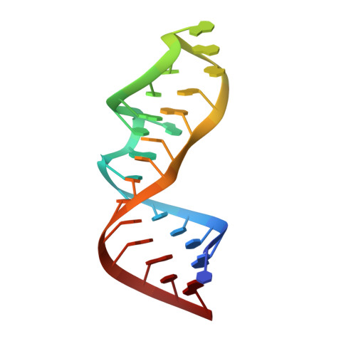
The ribose 2'-hydroxyl is the key chemical difference between RNA and DNA and primary source of their divergent structural and functional characteristics. Macromolecular X-ray diffraction experiments typically do not reveal the positions of hydrogen atoms. Thus, standard crystallography cannot determine 2'-OH orientation (H2'-C2'-O2'-HO2' torsion angle) and its potential roles in sculpting the RNA backbone and the expansive fold space. Here, we report the first neutron crystal structure of an RNA, the Escherichia coli rRNA Sarcin-Ricin Loop (SRL). 2'-OD orientations were established for all 27 residues and revealed O-D bonds pointing toward backbone (O3', 13 observations), nucleobase (11) or sugar (3). Most riboses in the SRL stem region show a 2'-OD backbone-orientation. GAGA-tetraloop riboses display a 2'-OD base-orientation. An atypical C2'-endo sugar pucker is strictly correlated with a 2'-OD sugar-orientation. Neutrons reveal the strong preference of the 2'-OH to donate in H-bonds and that 2'-OH orientation affects both backbone geometry and ribose pucker. We discuss 2'-OH and water molecule orientations in the SRL neutron structure and compare with results from a solution phase 10 μs MD simulation. We demonstrate that joint cryo-neutron/X-ray crystallography offers an all-in-one approach to determine the complete structural properties of RNA, i.e. geometry, conformation, protonation state and hydration structure.
- Department of Biochemistry, Vanderbilt University, Nashville, TN 37232, USA.
Organizational Affiliation: