7UB5 | pdb_00007ub5

SARS-CoV-2 Omicron-BA.2 3-RBD down Spike Protein Trimer without the P986-P987 stabilizing mutations (S-GSAS-Omicron-BA.2)


Experimental Data Snapshot

  • Method: ELECTRON MICROSCOPY
  • Resolution: 3.35 Å
  • Aggregation State: PARTICLE 
  • Reconstruction Method: SINGLE PARTICLE 

Starting Model: experimental
View more details

wwPDB Validation 3D Report Full Report

Validation slider image for 7UB5

This is version 1.5 of the entry. See complete history

Literature

Cryo-EM structures of SARS-CoV-2 Omicron BA.2 spike.

Stalls, V.Lindenberger, J.Gobeil, S.M.Henderson, R.Parks, R.Barr, M.Deyton, M.Martin, M.Janowska, K.Huang, X.May, A.Speakman, M.Beaudoin, E.Kraft, B.Lu, X.Edwards, R.J.Eaton, A.Montefiori, D.C.Williams, W.B.Saunders, K.O.Wiehe, K.Haynes, B.F.Acharya, P.

(2022) Cell Rep 39: 111009-111009

  • DOI: https://doi.org/10.1016/j.celrep.2022.111009
  • Primary Citation Related Structures: 
    7UB0, 7UB5, 7UB6

  • PubMed Abstract: 

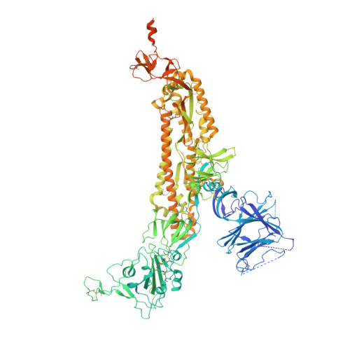
    The severe acute respiratory syndrome coronavirus 2 (SARS-CoV-2) Omicron BA.2 sub-lineage has gained in proportion relative to BA.1. Because spike (S) protein variations may underlie differences in their pathobiology, here we determine cryoelectron microscopy (cryo-EM) structures of the BA.2 S ectodomain and compare these with previously determined BA.1 S structures. BA.2 receptor-binding domain (RBD) mutations induce remodeling of the RBD structure, resulting in tighter packing and improved thermostability. Interprotomer RBD interactions are enhanced in the closed (or 3-RBD-down) BA.2 S, while the fusion peptide is less accessible to antibodies than in BA.1. Binding and pseudovirus neutralization assays reveal extensive immune evasion while defining epitopes of two outer RBD face-binding antibodies, DH1044 and DH1193, that neutralize both BA.1 and BA.2. Taken together, our results indicate that stabilization of the closed state through interprotomer RBD-RBD packing is a hallmark of the Omicron variant and show differences in key functional regions in the BA.1 and BA.2 S proteins.


  • Organizational Affiliation
    • Duke Human Vaccine Institute, Durham, NC 27710, USA.

Macromolecule Content 

  • Total Structure Weight: 437.56 kDa 
  • Atom Count: 24,923 
  • Modeled Residue Count: 3,104 
  • Deposited Residue Count: 3,855 
  • Unique protein chains: 1

Macromolecules

Find similar proteins by:|  3D Structure
Entity ID: 1
MoleculeChains  Sequence LengthOrganismDetailsImage
Spike glycoprotein
A, B, C
1,285Severe acute respiratory syndrome coronavirus 2Mutation(s): 23 
Gene Names: S2
UniProt
Find proteins for P0DTC2 (Severe acute respiratory syndrome coronavirus 2)
Explore P0DTC2 
Go to UniProtKB:  P0DTC2
Entity Groups
Sequence Clusters30% Identity50% Identity70% Identity90% Identity95% Identity100% Identity
UniProt GroupP0DTC2
Glycosylation
Glycosylation Sites: 11Go to GlyGen: P0DTC2-1
Sequence Annotations
Expand
Reference Sequence

Oligosaccharides

Help  
Entity ID: 2
MoleculeChains Length2D Diagram GlycosylationD Interactions
2-acetamido-2-deoxy-beta-D-glucopyranose-(1-4)-2-acetamido-2-deoxy-beta-D-glucopyranose
D,
E,
F,
G,
H,
D,
E,
F,
G,
H,
I,
J [auth K],
K [auth L],
L [auth M],
M [auth N]
2N-Glycosylation
Glycosylation Resources
GlyTouCan: G42666HT
GlyCosmos: G42666HT
GlyGen: G42666HT

Small Molecules

Ligands 1 Unique
IDChains Name / Formula / InChI Key2D Diagram3D Interactions
NAG

Query on NAG


Download:Ideal Coordinates CCD File
AA [auth B]
BA [auth B]
CA [auth B]
DA [auth B]
EA [auth B]
AA [auth B],
BA [auth B],
CA [auth B],
DA [auth B],
EA [auth B],
FA [auth B],
GA [auth C],
HA [auth C],
IA [auth C],
JA [auth C],
KA [auth C],
LA [auth C],
MA [auth C],
N [auth A],
NA [auth C],
O [auth A],
P [auth A],
Q [auth A],
R [auth A],
S [auth A],
T [auth A],
U [auth A],
V [auth A],
W [auth B],
X [auth B],
Y [auth B],
Z [auth B]
2-acetamido-2-deoxy-beta-D-glucopyranose
C8 H15 N O6
OVRNDRQMDRJTHS-FMDGEEDCSA-N

Experimental Data & Validation

Experimental Data

  • Method: ELECTRON MICROSCOPY
  • Resolution: 3.35 Å
  • Aggregation State: PARTICLE 
  • Reconstruction Method: SINGLE PARTICLE 
EM Software:
TaskSoftware PackageVersion
MODEL REFINEMENTPHENIX
RECONSTRUCTIONCoot
RECONSTRUCTIONISOLDE
RECONSTRUCTIONPHENIX

Structure Validation

View Full Validation Report


Entry History 

& Funding Information

Deposition Data


Funding OrganizationLocationGrant Number
National Institutes of Health/National Institute Of Allergy and Infectious Diseases (NIH/NIAID)United States--

Revision History  (Full details and data files)

  • Version 1.0: 2022-04-20
    Type: Initial release
  • Version 1.1: 2022-04-27
    Changes: Database references, Source and taxonomy
  • Version 1.2: 2022-07-06
    Changes: Database references
  • Version 1.3: 2022-07-13
    Changes: Database references
  • Version 1.4: 2024-10-23
    Changes: Data collection, Refinement description, Structure summary
  • Version 1.5: 2025-05-21
    Changes: Data collection