7UA3 | pdb_00007ua3

Structure of PKA phosphorylated human RyR2-R2474S in the closed state in the presence of Calmodulin


Experimental Data Snapshot

  • Method: ELECTRON MICROSCOPY
  • Resolution: 2.97 Å
  • Aggregation State: PARTICLE 
  • Reconstruction Method: SINGLE PARTICLE 

Starting Model: experimental
View more details

wwPDB Validation 3D Report Full Report

Validation slider image for 7UA3

This is version 1.1 of the entry. See complete history

Literature

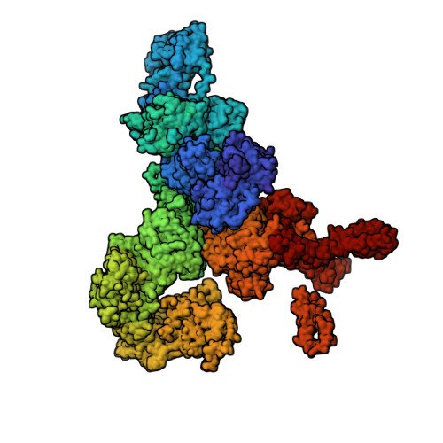
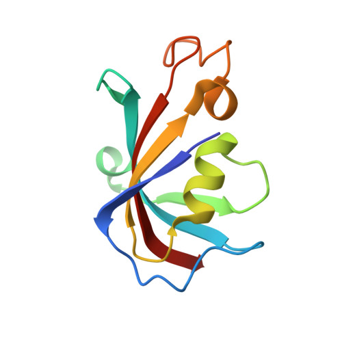
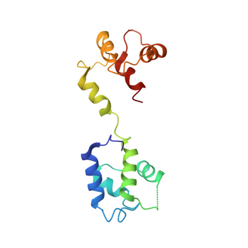
Structural analyses of human ryanodine receptor type 2 channels reveal the mechanisms for sudden cardiac death and treatment.

Miotto, M.C.Weninger, G.Dridi, H.Yuan, Q.Liu, Y.Wronska, A.Melville, Z.Sittenfeld, L.Reiken, S.Marks, A.R.

(2022) Sci Adv 8: eabo1272-eabo1272

  • DOI: https://doi.org/10.1126/sciadv.abo1272
  • Primary Citation Related Structures: 
    7U9Q, 7U9R, 7U9T, 7U9X, 7U9Z, 7UA1, 7UA3, 7UA4, 7UA5, 7UA9

  • PubMed Abstract: 

    Ryanodine receptor type 2 (RyR2) mutations have been linked to an inherited form of exercise-induced sudden cardiac death called catecholaminergic polymorphic ventricular tachycardia (CPVT). CPVT results from stress-induced sarcoplasmic reticular Ca 2+ leak via the mutant RyR2 channels during diastole. We present atomic models of human wild-type (WT) RyR2 and the CPVT mutant RyR2-R2474S determined by cryo-electron microscopy with overall resolutions in the range of 2.6 to 3.6 Å, and reaching local resolutions of 2.25 Å, unprecedented for RyR2 channels. Under nonactivating conditions, the RyR2-R2474S channel is in a "primed" state between the closed and open states of WT RyR2, rendering it more sensitive to activation that results in stress-induced Ca 2+ leak. The Rycal drug ARM210 binds to RyR2-R2474S, reverting the primed state toward the closed state. Together, these studies provide a mechanism for CPVT and for the therapeutic actions of ARM210.


  • Organizational Affiliation
    • Department of Physiology and Cellular Biophysics, Columbia University Vagelos College of Physicians and Surgeons, New York, NY, USA.

Macromolecule Content 

  • Total Structure Weight: 2,379.79 kDa 
  • Atom Count: 150,252 
  • Modeled Residue Count: 18,768 
  • Deposited Residue Count: 20,896 
  • Unique protein chains: 3

Macromolecules

Find similar proteins by:|  3D Structure
Entity ID: 1
MoleculeChains  Sequence LengthOrganismDetailsImage
Ryanodine receptor 2A,
J [auth B],
K [auth C],
L [auth D]
4,967Homo sapiensMutation(s): 1 
Gene Names: RYR2
Membrane Entity: Yes 
UniProt & NIH Common Fund Data Resources
Find proteins for Q92736 (Homo sapiens)
Explore Q92736 
Go to UniProtKB:  Q92736
PHAROS:  Q92736
GTEx:  ENSG00000198626 
Entity Groups
Sequence Clusters30% Identity50% Identity70% Identity90% Identity95% Identity100% Identity
UniProt GroupQ92736
Sequence Annotations
Expand
Reference Sequence
Find similar proteins by:|  3D Structure
Entity ID: 2
MoleculeChains  Sequence LengthOrganismDetailsImage
Peptidyl-prolyl cis-trans isomerase FKBP1BB [auth E],
C [auth F],
D [auth G],
E [auth H]
108Homo sapiensMutation(s): 0 
Gene Names: FKBP1BFKBP12.6FKBP1LFKBP9OTK4
EC: 5.2.1.8
UniProt & NIH Common Fund Data Resources
Find proteins for P68106 (Homo sapiens)
Explore P68106 
Go to UniProtKB:  P68106
PHAROS:  P68106
GTEx:  ENSG00000119782 
Entity Groups
Sequence Clusters30% Identity50% Identity70% Identity90% Identity95% Identity100% Identity
UniProt GroupP68106
Sequence Annotations
Expand
Reference Sequence
Find similar proteins by:|  3D Structure
Entity ID: 3
MoleculeChains  Sequence LengthOrganismDetailsImage
Calmodulin-1F [auth I],
G [auth J],
H [auth K],
I [auth L]
149Homo sapiensMutation(s): 0 
Gene Names: CALM1CALMCAMCAM1
UniProt & NIH Common Fund Data Resources
Find proteins for P0DP23 (Homo sapiens)
Explore P0DP23 
Go to UniProtKB:  P0DP23
PHAROS:  P0DP23
GTEx:  ENSG00000198668 
Entity Groups
Sequence Clusters30% Identity50% Identity70% Identity90% Identity95% Identity100% Identity
UniProt GroupP0DP23
Sequence Annotations
Expand
Reference Sequence

Small Molecules

Ligands 2 Unique
IDChains Name / Formula / InChI Key2D Diagram3D Interactions
ATP
(Subject of Investigation/LOI)

Query on ATP


Download:Ideal Coordinates CCD File
N [auth A]
O [auth A]
Q [auth B]
R [auth B]
T [auth C]
N [auth A],
O [auth A],
Q [auth B],
R [auth B],
T [auth C],
U [auth C],
W [auth D],
X [auth D]
ADENOSINE-5'-TRIPHOSPHATE
C10 H16 N5 O13 P3
ZKHQWZAMYRWXGA-KQYNXXCUSA-N
ZN
(Subject of Investigation/LOI)

Query on ZN


Download:Ideal Coordinates CCD File
M [auth A],
P [auth B],
S [auth C],
V [auth D]
ZINC ION
Zn
PTFCDOFLOPIGGS-UHFFFAOYSA-N

Experimental Data & Validation

Experimental Data

  • Method: ELECTRON MICROSCOPY
  • Resolution: 2.97 Å
  • Aggregation State: PARTICLE 
  • Reconstruction Method: SINGLE PARTICLE 

Structure Validation

View Full Validation Report


Entry History 

& Funding Information

Deposition Data


Funding OrganizationLocationGrant Number
National Institutes of Health/National Heart, Lung, and Blood Institute (NIH/NHLBI)United StatesR01HL145473

Revision History  (Full details and data files)

  • Version 1.0: 2022-08-03
    Type: Initial release
  • Version 1.1: 2024-11-06
    Changes: Data collection, Refinement description, Structure summary