7U32 | pdb_00007u32

MVV cleaved synaptic complex (CSC) intasome at 3.4 A resolution


Experimental Data Snapshot

  • Method: ELECTRON MICROSCOPY
  • Resolution: 3.46 Å
  • Aggregation State: PARTICLE 
  • Reconstruction Method: SINGLE PARTICLE 

wwPDB Validation 3D Report Full Report

Validation slider image for 7U32

This is version 1.2 of the entry. See complete history

Literature

Multivalent interactions essential for lentiviral integrase function.

Ballandras-Colas, A.Chivukula, V.Gruszka, D.T.Shan, Z.Singh, P.K.Pye, V.E.McLean, R.K.Bedwell, G.J.Li, W.Nans, A.Cook, N.J.Fadel, H.J.Poeschla, E.M.Griffiths, D.J.Vargas, J.Taylor, I.A.Lyumkis, D.Yardimci, H.Engelman, A.N.Cherepanov, P.

(2022) Nat Commun 13: 2416-2416

  • DOI: https://doi.org/10.1038/s41467-022-29928-8
  • Primary Citation Related Structures: 
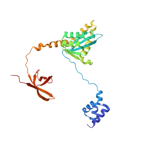
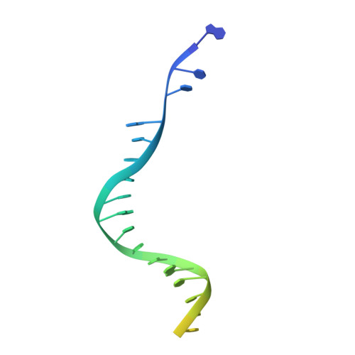
    7U32

  • PubMed Abstract: 

    A multimer of retroviral integrase (IN) synapses viral DNA ends within a stable intasome nucleoprotein complex for integration into a host cell genome. Reconstitution of the intasome from the maedi-visna virus (MVV), an ovine lentivirus, revealed a large assembly containing sixteen IN subunits 1 . Herein, we report cryo-EM structures of the lentiviral intasome prior to engagement of target DNA and following strand transfer, refined at 3.4 and 3.5 Å resolution, respectively. The structures elucidate details of the protein-protein and protein-DNA interfaces involved in lentiviral intasome formation. We show that the homomeric interfaces involved in IN hexadecamer formation and the α-helical configuration of the linker connecting the C-terminal and catalytic core domains are critical for MVV IN strand transfer activity in vitro and for virus infectivity. Single-molecule microscopy in conjunction with photobleaching reveals that the MVV intasome can bind a variable number, up to sixteen molecules, of the lentivirus-specific host factor LEDGF/p75. Concordantly, ablation of endogenous LEDGF/p75 results in gross redistribution of MVV integration sites in human and ovine cells. Our data confirm the importance of the expanded architecture observed in cryo-EM studies of lentiviral intasomes and suggest that this organization underlies multivalent interactions with chromatin for integration targeting to active genes.


  • Organizational Affiliation
    • Chromatin Structure and Mobile DNA Laboratory, The Francis Crick Institute, London, UK.

Macromolecule Content 

  • Total Structure Weight: 553.2 kDa 
  • Atom Count: 32,150 
  • Modeled Residue Count: 3,866 
  • Deposited Residue Count: 4,608 
  • Unique protein chains: 1
  • Unique nucleic acid chains: 2

Macromolecules


Find similar proteins by:|  3D Structure
Entity ID: 1
MoleculeChains  Sequence LengthOrganismDetailsImage
Integrase281Visna/maedi virus EV1 KV1772Mutation(s): 0 
Gene Names: pol
EC: 2.7.7 (PDB Primary Data), 3.1 (PDB Primary Data)
UniProt
Find proteins for P35956 (Maedi visna virus (strain KV1772))
Explore P35956 
Go to UniProtKB:  P35956
Entity Groups
Sequence Clusters30% Identity50% Identity70% Identity90% Identity95% Identity100% Identity
UniProt GroupP35956
Sequence Annotations
Expand
Reference Sequence
Find similar nucleic acids by:  (by identity cutoff) 
Entity ID: 2
MoleculeChains LengthOrganismImage
DNA EV273I [auth X],
S [auth Y]
29synthetic construct
Sequence Annotations
Expand
Reference Sequence
Find similar nucleic acids by:  (by identity cutoff) 
Entity ID: 3
MoleculeChains LengthOrganismImage
DNA EV272J [auth W],
T [auth Z]
27synthetic construct
Sequence Annotations
Expand
Reference Sequence

Small Molecules

Ligands 2 Unique
IDChains Name / Formula / InChI Key2D Diagram3D Interactions
ZN

Query on ZN


Download:Ideal Coordinates CCD File
AA [auth G]
BA [auth I]
DA [auth J]
EA [auth K]
FA [auth M]
AA [auth G],
BA [auth I],
DA [auth J],
EA [auth K],
FA [auth M],
GA [auth N],
HA [auth O],
U [auth A],
W [auth B],
X [auth C],
Y [auth E],
Z [auth F]
ZINC ION
Zn
PTFCDOFLOPIGGS-UHFFFAOYSA-N
CA

Query on CA


Download:Ideal Coordinates CCD File
CA [auth I],
V [auth A]
CALCIUM ION
Ca
BHPQYMZQTOCNFJ-UHFFFAOYSA-N

Experimental Data & Validation

Experimental Data

  • Method: ELECTRON MICROSCOPY
  • Resolution: 3.46 Å
  • Aggregation State: PARTICLE 
  • Reconstruction Method: SINGLE PARTICLE 
EM Software:
TaskSoftware PackageVersion
RECONSTRUCTIONcryoSPARC3.0
MODEL REFINEMENTCoot
MODEL REFINEMENTPHENIX

Structure Validation

View Full Validation Report


Entry History 

& Funding Information

Deposition Data


Funding OrganizationLocationGrant Number
National Science Foundation (NSF, United States)United StatesMCB-2048095
National Institutes of Health/National Institute Of Allergy and Infectious Diseases (NIH/NIAID)United StatesU54 AI150472
National Institutes of Health/National Institute Of Allergy and Infectious Diseases (NIH/NIAID)United StatesR01 AI136680
The Francis Crick InstituteUnited KingdomFC10061
National Institutes of Health/National Institute Of Allergy and Infectious Diseases (NIH/NIAID)United StatesP50AI150481

Revision History  (Full details and data files)

  • Version 1.0: 2022-05-11
    Type: Initial release
  • Version 1.1: 2024-02-21
    Changes: Data collection
  • Version 1.2: 2025-05-28
    Changes: Data collection, Structure summary