7TXL

Crystal structure of EgtU solute binding domain from Streptococcus pneumoniae D39 in complex with L-ergothioneine


Experimental Data Snapshot

  • Method: X-RAY DIFFRACTION
  • Resolution: 2.44 Å
  • R-Value Free: 0.248 
  • R-Value Work: 0.196 
  • R-Value Observed: 0.198 

wwPDB Validation   3D Report Full Report


Ligand Structure Quality Assessment 


This is version 1.1 of the entry. See complete history


Literature

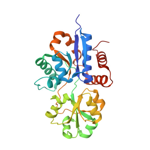
Discovery and structure of a widespread bacterial ABC transporter specific for ergothioneine.

Zhang, Y.Gonzalez-Gutierrez, G.Legg, K.A.Walsh, B.J.C.Pis Diez, C.M.Edmonds, K.A.Giedroc, D.P.

(2022) Nat Commun 13: 7586-7586

  • DOI: https://doi.org/10.1038/s41467-022-35277-3
  • Primary Citation of Related Structures:  
    7TXK, 7TXL

  • PubMed Abstract: 

    L-Ergothioneine (ET), the 2-thioimidazole derivative of trimethylhistidine, is biosynthesized by select fungi and bacteria, notably Mycobacterium tuberculosis, and functions as a scavenger of reactive oxygen species. The extent to which ET broadly functions in bacterial cells unable to synthesize it is unknown. Here we show that spd_1642-1643 in Streptococcus pneumoniae, a Gram-positive respiratory pathogen, encodes an ET uptake ATP-binding cassette (ABC) transporter, designated EgtU. The solute binding domain (SBD) of EgtU, EgtUC, binds ET with high affinity and exquisite specificity in a cleft between the two subdomains, with cation-π interactions engaging the betaine moiety and a network of water molecules that surround the thioimidazole ring. EgtU is highly conserved among known quaternary amine compound-specific transporters and widely distributed in Firmicutes, including the human pathogens Listeria monocytogenes, as BilEB, Enterococcus faecalis and Staphylococcus aureus. ET increases the chemical diversity of the low molecular weight thiol pool in Gram-positive human pathogens and may contribute to antioxidant defenses in the infected host.


  • Organizational Affiliation

    Department of Chemistry, Indiana University, Bloomington, IN, 47405-7102, USA.


Macromolecules
Find similar proteins by:  (by identity cutoff)  |  3D Structure
Entity ID: 1
MoleculeChains Sequence LengthOrganismDetailsImage
Choline transporter (Glycine betaine transport system permease protein)
A, B
277Streptococcus pneumoniae D39Mutation(s): 0 
Gene Names: proWXSPD_1642
UniProt
Find proteins for A0A0H2ZQB9 (Streptococcus pneumoniae serotype 2 (strain D39 / NCTC 7466))
Explore A0A0H2ZQB9 
Go to UniProtKB:  A0A0H2ZQB9
Entity Groups  
Sequence Clusters30% Identity50% Identity70% Identity90% Identity95% Identity100% Identity
UniProt GroupA0A0H2ZQB9
Sequence Annotations
Expand
  • Reference Sequence
Small Molecules
Ligands 2 Unique
IDChains Name / Formula / InChI Key2D Diagram3D Interactions
LW8 (Subject of Investigation/LOI)
Query on LW8

Download Ideal Coordinates CCD File 
C [auth A],
E [auth B]
trimethyl-[(2S)-1-oxidanyl-1-oxidanylidene-3-(2-sulfanylidene-1,3-dihydroimidazol-4-yl)propan-2-yl]azanium
C9 H16 N3 O2 S
SSISHJJTAXXQAX-ZETCQYMHSA-O
EDO
Query on EDO

Download Ideal Coordinates CCD File 
D [auth A]1,2-ETHANEDIOL
C2 H6 O2
LYCAIKOWRPUZTN-UHFFFAOYSA-N
Experimental Data & Validation

Experimental Data

  • Method: X-RAY DIFFRACTION
  • Resolution: 2.44 Å
  • R-Value Free: 0.248 
  • R-Value Work: 0.196 
  • R-Value Observed: 0.198 
  • Space Group: F 2 2 2
Unit Cell:
Length ( Å )Angle ( ˚ )
a = 119.868α = 90
b = 128.743β = 90
c = 207.54γ = 90
Software Package:
Software NamePurpose
PHENIXrefinement
XDSdata reduction
XDSdata scaling
PHASERphasing

Structure Validation

View Full Validation Report


Ligand Structure Quality Assessment 


Entry History & Funding Information

Deposition Data


Funding OrganizationLocationGrant Number
National Institutes of Health/National Institute of General Medical Sciences (NIH/NIGMS)United StatesR35 GM118157

Revision History  (Full details and data files)

  • Version 1.0: 2022-12-21
    Type: Initial release
  • Version 1.1: 2023-10-25
    Changes: Data collection, Refinement description