7TW1

Cryo-EM structure of human band 3-protein 4.2 complex (B2P2vertical)


Experimental Data Snapshot

  • Method: ELECTRON MICROSCOPY
  • Resolution: 4.60 Å
  • Aggregation State: PARTICLE 
  • Reconstruction Method: SINGLE PARTICLE 

wwPDB Validation   3D Report Full Report


This is version 1.3 of the entry. See complete history


Literature

Structure, dynamics and assembly of the ankyrin complex on human red blood cell membrane.

Xia, X.Liu, S.Zhou, Z.H.

(2022) Nat Struct Mol Biol 29: 698-705

  • DOI: https://doi.org/10.1038/s41594-022-00779-7
  • Primary Citation of Related Structures:  
    7TVZ, 7TW0, 7TW1, 7TW2, 7TW3, 7TW5, 7TW6

  • PubMed Abstract: 

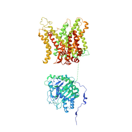
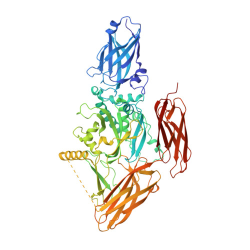
    The cytoskeleton of a red blood cell (RBC) is anchored to the cell membrane by the ankyrin complex. This complex is assembled during RBC genesis and comprises primarily band 3, protein 4.2 and ankyrin, whose mutations contribute to numerous human inherited diseases. High-resolution structures of the ankyrin complex have been long sought-after to understand its assembly and disease-causing mutations. Here, we analyzed native complexes on the human RBC membrane by stepwise fractionation. Cryo-electron microscopy structures of nine band-3-associated complexes reveal that protein 4.2 stabilizes the cytoplasmic domain of band 3 dimer. In turn, the superhelix-shaped ankyrin binds to this protein 4.2 via ankyrin repeats (ARs) 6-13 and to another band 3 dimer via ARs 17-20, bridging two band 3 dimers in the ankyrin complex. Integration of these structures with both prior data and our biochemical data supports a model of ankyrin complex assembly during erythropoiesis and identifies interactions essential for the mechanical stability of RBC.


  • Organizational Affiliation

    Department of Microbiology, Immunology, and Molecular Genetics, University of California, Los Angeles, CA, USA.


Macromolecules
Find similar proteins by:  (by identity cutoff)  |  3D Structure
Entity ID: 1
MoleculeChains Sequence LengthOrganismDetailsImage
Band 3 anion transport proteinA [auth B],
B [auth A]
911Homo sapiensMutation(s): 0 
Membrane Entity: Yes 
UniProt & NIH Common Fund Data Resources
Find proteins for P02730 (Homo sapiens)
Explore P02730 
Go to UniProtKB:  P02730
PHAROS:  P02730
GTEx:  ENSG00000004939 
Entity Groups  
Sequence Clusters30% Identity50% Identity70% Identity90% Identity95% Identity100% Identity
UniProt GroupP02730
Sequence Annotations
Expand
  • Reference Sequence
Find similar proteins by:  (by identity cutoff)  |  3D Structure
Entity ID: 2
MoleculeChains Sequence LengthOrganismDetailsImage
Protein 4.2C [auth E],
D [auth F]
691Homo sapiensMutation(s): 0 
Membrane Entity: Yes 
UniProt & NIH Common Fund Data Resources
Find proteins for P16452 (Homo sapiens)
Explore P16452 
Go to UniProtKB:  P16452
PHAROS:  P16452
GTEx:  ENSG00000166947 
Entity Groups  
Sequence Clusters30% Identity50% Identity70% Identity90% Identity95% Identity100% Identity
UniProt GroupP16452
Sequence Annotations
Expand
  • Reference Sequence
Experimental Data & Validation

Experimental Data

  • Method: ELECTRON MICROSCOPY
  • Resolution: 4.60 Å
  • Aggregation State: PARTICLE 
  • Reconstruction Method: SINGLE PARTICLE 
EM Software:
TaskSoftware PackageVersion
RECONSTRUCTIONRELION3.1
MODEL REFINEMENTPHENIX1.19

Structure Validation

View Full Validation Report


Entry History & Funding Information

Deposition Data


Funding OrganizationLocationGrant Number
National Institutes of Health/National Heart, Lung, and Blood Institute (NIH/NHLBI)United StatesR01GM071940

Revision History  (Full details and data files)

  • Version 1.0: 2022-06-08
    Type: Initial release
  • Version 1.1: 2022-06-22
    Changes: Database references
  • Version 1.2: 2022-07-27
    Changes: Database references
  • Version 1.3: 2024-02-21
    Changes: Data collection