7TIF | pdb_00007tif

2.85 Angstroem crystal structure of Arginyltransferase 1 (ATE1) from Saccharomyces cerevisiae


Experimental Data Snapshot

  • Method: X-RAY DIFFRACTION
  • Resolution: 2.85 Å
  • R-Value Free: 
    0.257 (Depositor), 0.247 (DCC) 
  • R-Value Work: 
    0.200 (Depositor) 
  • R-Value Observed: 
    0.203 (Depositor) 

Starting Model: in silico
View more details

wwPDB Validation 3D Report Full Report

Validation slider image for 7TIF

This is version 1.3 of the entry. See complete history

Literature

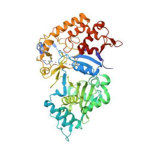
The Structure of Saccharomyces cerevisiae Arginyltransferase 1 (ATE1).

Van, V.Ejimogu, N.E.Bui, T.S.Smith, A.T.

(2022) J Mol Biology 434: 167816-167816

  • DOI: https://doi.org/10.1016/j.jmb.2022.167816
  • Primary Citation Related Structures: 
    7TIF

  • PubMed Abstract: 

    Eukaryotic post-translational arginylation, mediated by the family of enzymes known as the arginyltransferases (ATE1s), is an important post-translational modification that can alter protein function and even dictate cellular protein half-life. Multiple major biological pathways are linked to the fidelity of this process, including neural and cardiovascular developments, cell division, and even the stress response. Despite this significance, the structural, mechanistic, and regulatory mechanisms that govern ATE1 function remain enigmatic. To that end, we have used X-ray crystallography to solve the crystal structure of ATE1 from the model organism Saccharomyces cerevisiae ATE1 (ScATE1) in the apo form. The three-dimensional structure of ScATE1 reveals a bilobed protein containing a GCN5-related N-acetyltransferase (GNAT) fold, and this crystalline behavior is faithfully recapitulated in solution based on size-exclusion chromatography-coupled small angle X-ray scattering (SEC-SAXS) analyses and cryo-EM 2D class averaging. Structural superpositions and electrostatic analyses point to this domain and its domain-domain interface as the location of catalytic activity and tRNA binding, and these comparisons strongly suggest a mechanism for post-translational arginylation. Additionally, our structure reveals that the N-terminal domain, which we have previously shown to bind a regulatory [Fe-S] cluster, is dynamic and disordered in the absence of metal bound in this location, hinting at the regulatory influence of this region. When taken together, these insights bring us closer to answering pressing questions regarding the molecular-level mechanism of eukaryotic post-translational arginylation.


  • Organizational Affiliation
    • Department of Chemistry and Biochemistry, University of Maryland, Baltimore County, Baltimore, MD 21250, USA. Electronic address: https://twitter.com/VernaVan.

Macromolecule Content 

  • Total Structure Weight: 742.73 kDa 
  • Atom Count: 46,039 
  • Modeled Residue Count: 5,493 
  • Deposited Residue Count: 6,216 
  • Unique protein chains: 1

Macromolecules

Find similar proteins by:|  3D Structure
Entity ID: 1
MoleculeChains  Sequence LengthOrganismDetailsImage
Arginyl-tRNA--protein transferase 1
A, B, C, D, E
A, B, C, D, E, F, G, H, I, J, K, L
518Saccharomyces cerevisiaeMutation(s): 0 
Gene Names: ATE1YGL017W
EC: 2.3.2.8
UniProt
Find proteins for P16639 (Saccharomyces cerevisiae (strain ATCC 204508 / S288c))
Explore P16639 
Go to UniProtKB:  P16639
Entity Groups
Sequence Clusters30% Identity50% Identity70% Identity90% Identity95% Identity100% Identity
UniProt GroupP16639
Sequence Annotations
Expand
Reference Sequence

Small Molecules

Ligands 2 Unique
IDChains Name / Formula / InChI Key2D Diagram3D Interactions
SO4

Query on SO4


Download:Ideal Coordinates CCD File
AA [auth A]
AB [auth B]
AC [auth C]
AD [auth D]
AE [auth E]
AA [auth A],
AB [auth B],
AC [auth C],
AD [auth D],
AE [auth E],
AF [auth F],
AG [auth G],
AH [auth I],
AI [auth K],
BA [auth A],
BB [auth B],
BC [auth C],
BD [auth D],
BE [auth E],
BF [auth F],
BG [auth G],
BH [auth I],
BI [auth K],
CA [auth A],
CB [auth B],
CC [auth C],
CD [auth D],
CE [auth E],
CF [auth F],
CG [auth G],
CH [auth I],
CI [auth K],
DA [auth A],
DB [auth B],
DC [auth C],
DD [auth D],
DE [auth E],
DF [auth F],
DH [auth I],
DI [auth K],
EA [auth A],
EB [auth B],
EC [auth C],
ED [auth D],
EE [auth E],
EF [auth F],
EH [auth I],
EI [auth K],
FA [auth A],
FB [auth B],
FC [auth C],
FD [auth D],
FE [auth E],
FF [auth F],
FH [auth I],
FI [auth K],
GA [auth A],
GB [auth B],
GC [auth C],
GD [auth D],
GE [auth E],
GF [auth F],
GH [auth I],
GI [auth K],
HA [auth A],
HB [auth B],
HC [auth C],
HD [auth D],
HE [auth E],
HF [auth F],
HH [auth I],
IA [auth A],
IB [auth B],
IC [auth C],
ID [auth D],
IE [auth E],
IF [auth F],
IH [auth I],
JB [auth B],
JC [auth C],
JD [auth D],
JE [auth E],
JF [auth F],
JG [auth H],
JH [auth I],
KB [auth B],
KC [auth C],
KD [auth D],
KE [auth E],
KG [auth H],
KH [auth I],
KI [auth L],
LB [auth B],
LC [auth C],
LD [auth D],
LE [auth E],
LG [auth H],
LH [auth I],
LI [auth L],
M [auth A],
MB [auth B],
MC [auth C],
MD [auth D],
ME [auth E],
MF [auth G],
MG [auth H],
MH [auth I],
MI [auth L],
N [auth A],
NB [auth B],
NC [auth C],
ND [auth D],
NE [auth E],
NF [auth G],
NG [auth H],
NI [auth L],
O [auth A],
OB [auth B],
OC [auth C],
OD [auth D],
OE [auth E],
OF [auth G],
OG [auth H],
OH [auth J],
OI [auth L],
P [auth A],
PB [auth B],
PC [auth C],
PD [auth D],
PE [auth E],
PF [auth G],
PG [auth H],
PH [auth J],
PI [auth L],
Q [auth A],
QC [auth C],
QD [auth D],
QE [auth E],
QF [auth G],
QG [auth H],
QH [auth J],
QI [auth L],
R [auth A],
RA [auth B],
RC [auth C],
RE [auth E],
RF [auth G],
RG [auth H],
RH [auth J],
RI [auth L],
S [auth A],
SA [auth B],
SE [auth E],
SF [auth G],
SG [auth H],
SH [auth J],
SI [auth L],
T [auth A],
TA [auth B],
TE [auth E],
TF [auth G],
TG [auth H],
TH [auth J],
TI [auth L],
U [auth A],
UA [auth B],
UF [auth G],
UG [auth H],
UH [auth J],
UI [auth L],
V [auth A],
VA [auth B],
VB [auth C],
VC [auth D],
VD [auth E],
VF [auth G],
VG [auth H],
VH [auth J],
W [auth A],
WA [auth B],
WB [auth C],
WC [auth D],
WD [auth E],
WF [auth G],
X [auth A],
XA [auth B],
XB [auth C],
XC [auth D],
XD [auth E],
XE [auth F],
XF [auth G],
XH [auth K],
Y [auth A],
YA [auth B],
YB [auth C],
YC [auth D],
YD [auth E],
YE [auth F],
YF [auth G],
YH [auth K],
Z [auth A],
ZA [auth B],
ZB [auth C],
ZC [auth D],
ZD [auth E],
ZE [auth F],
ZF [auth G],
ZH [auth K]
SULFATE ION
O4 S
QAOWNCQODCNURD-UHFFFAOYSA-L
GOL

Query on GOL


Download:Ideal Coordinates CCD File
DG [auth G]
EG [auth G]
FG [auth G]
GG [auth G]
HG [auth G]
DG [auth G],
EG [auth G],
FG [auth G],
GG [auth G],
HG [auth G],
HI [auth K],
IG [auth G],
II [auth K],
JA [auth A],
JI [auth K],
KA [auth A],
KF [auth F],
LA [auth A],
LF [auth F],
MA [auth A],
NA [auth A],
NH [auth I],
OA [auth A],
PA [auth A],
QA [auth A],
QB [auth B],
RB [auth B],
RD [auth D],
SB [auth B],
SC [auth C],
SD [auth D],
TB [auth B],
TC [auth C],
TD [auth D],
UB [auth B],
UC [auth C],
UD [auth D],
UE [auth E],
VE [auth E],
VI [auth L],
WE [auth E],
WG [auth H],
WH [auth J],
XG [auth H],
YG [auth H],
ZG [auth H]
GLYCEROL
C3 H8 O3
PEDCQBHIVMGVHV-UHFFFAOYSA-N

Experimental Data & Validation

Experimental Data

  • Method: X-RAY DIFFRACTION
  • Resolution: 2.85 Å
  • R-Value Free:  0.257 (Depositor), 0.247 (DCC) 
  • R-Value Work:  0.200 (Depositor) 
  • R-Value Observed: 0.203 (Depositor) 
Space Group: P 43
Unit Cell:
Length ( Å )Angle ( ˚ )
a = 235.27α = 90
b = 235.27β = 90
c = 171.1γ = 90
Software Package:
Software NamePurpose
PHENIXrefinement
xia2data reduction
xia2data scaling
PHASERphasing

Structure Validation

View Full Validation Report


Entry History 

& Funding Information

Deposition Data


Funding OrganizationLocationGrant Number
National Institutes of Health/National Institute of General Medical Sciences (NIH/NIGMS)United StatesR35GM133497

Revision History  (Full details and data files)

  • Version 1.0: 2022-09-14
    Type: Initial release
  • Version 1.1: 2022-09-21
    Changes: Database references
  • Version 1.2: 2022-10-05
    Changes: Database references
  • Version 1.3: 2024-04-03
    Changes: Data collection, Refinement description