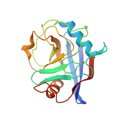
7THD

Structure of Cyclophilin D Peptidyl-Prolyl Isomerase Domain bound to Macrocyclic Inhibitor B52

  • Classification: ISOMERASE
  • Organism(s): Homo sapiens
  • Expression System: Escherichia coli BL21(DE3)
  • Mutation(s): Yes 

  • Deposited: 2022-01-10 Released: 2022-08-24 
  • Deposition Author(s): Rangwala, A.M., Thakur, M.K., Seeliger, M.A., Peterson, A.A., Liu, D.R.
  • Funding Organization(s): National Institutes of Health/National Cancer Institute (NIH/NCI), National Institutes of Health/National Institute of General Medical Sciences (NIH/NIGMS), National Institutes of Health/National Institute of Biomedical Imaging and Bioengineering (NIH/NIBIB), National Institutes of Health/Office of the Director, Howard Hughes Medical Institute (HHMI)

Experimental Data Snapshot

  • Method: X-RAY DIFFRACTION
  • Resolution: 1.16 Å
  • R-Value Free: 0.152 
  • R-Value Work: 0.136 
  • R-Value Observed: 0.136 

wwPDB Validation   3D Report Full Report


Ligand Structure Quality Assessment 


This is version 1.3 of the entry. See complete history


Literature

Discovery and molecular basis of subtype-selective cyclophilin inhibitors.

Peterson, A.A.Rangwala, A.M.Thakur, M.K.Ward, P.S.Hung, C.Outhwaite, I.R.Chan, A.I.Usanov, D.L.Mootha, V.K.Seeliger, M.A.Liu, D.R.

(2022) Nat Chem Biol 18: 1184-1195

  • DOI: https://doi.org/10.1038/s41589-022-01116-1
  • Primary Citation of Related Structures:  
    7TGS, 7TGT, 7TGU, 7TGV, 7TH1, 7TH6, 7TH7, 7THC, 7THD, 7THF

  • PubMed Abstract: 

    Although cyclophilins are attractive targets for probing biology and therapeutic intervention, no subtype-selective cyclophilin inhibitors have been described. We discovered novel cyclophilin inhibitors from the in vitro selection of a DNA-templated library of 256,000 drug-like macrocycles for cyclophilin D (CypD) affinity. Iterated macrocycle engineering guided by ten X-ray co-crystal structures yielded potent and selective inhibitors (half maximal inhibitory concentration (IC 50 ) = 10 nM) that bind the active site of CypD and also make novel interactions with non-conserved residues in the S2 pocket, an adjacent exo-site. The resulting macrocycles inhibit CypD activity with 21- to >10,000-fold selectivity over other cyclophilins and inhibit mitochondrial permeability transition pore opening in isolated mitochondria. We further exploited S2 pocket interactions to develop the first cyclophilin E (CypE)-selective inhibitor, which forms a reversible covalent bond with a CypE S2 pocket lysine, and exhibits 30- to >4,000-fold selectivity over other cyclophilins. These findings reveal a strategy to generate isoform-selective small-molecule cyclophilin modulators, advancing their suitability as targets for biological investigation and therapeutic development.


  • Organizational Affiliation

    Merkin Institute of Transformative Technologies in Healthcare, Broad Institute of MIT and Harvard, Cambridge, MA, USA.


Macromolecules
Find similar proteins by:  (by identity cutoff)  |  3D Structure
Entity ID: 1
MoleculeChains Sequence LengthOrganismDetailsImage
Peptidyl-prolyl cis-trans isomerase F, mitochondrial163Homo sapiensMutation(s): 1 
Gene Names: PPIFCYP3
EC: 5.2.1.8
UniProt & NIH Common Fund Data Resources
Find proteins for P30405 (Homo sapiens)
Explore P30405 
Go to UniProtKB:  P30405
PHAROS:  P30405
GTEx:  ENSG00000108179 
Entity Groups  
Sequence Clusters30% Identity50% Identity70% Identity90% Identity95% Identity100% Identity
UniProt GroupP30405
Sequence Annotations
Expand
  • Reference Sequence
Small Molecules
Ligands 1 Unique
IDChains Name / Formula / InChI Key2D Diagram3D Interactions
I4V (Subject of Investigation/LOI)
Query on I4V

Download Ideal Coordinates CCD File 
B [auth A][(4'-{[(4S,7S,11R,13E,19S)-19-{[2-(2-aminoethoxy)ethyl]carbamoyl}-7-benzyl-3,6,12,15,21-pentaoxo-1,3,4,5,6,7,8,9,10,12,15,16,17,18,19,20,21,22-octadecahydro-2H-7,11-methano-2,5,11,16,20-benzopentaazacyclotetracosin-4-yl]methyl}[1,1'-biphenyl]-4-yl)methyl]propanedioic acid
C53 H61 N7 O11
ZHNYUSCORILKFS-FCZRSVTRSA-N
Experimental Data & Validation

Experimental Data

  • Method: X-RAY DIFFRACTION
  • Resolution: 1.16 Å
  • R-Value Free: 0.152 
  • R-Value Work: 0.136 
  • R-Value Observed: 0.136 
  • Space Group: P 2 21 21
Unit Cell:
Length ( Å )Angle ( ˚ )
a = 38.408α = 90
b = 67.032β = 90
c = 69.324γ = 90
Software Package:
Software NamePurpose
PHENIXrefinement
Cootmodel building
XDSdata reduction
autoPROCdata scaling
autoPROCdata processing
PHASERphasing

Structure Validation

View Full Validation Report


Ligand Structure Quality Assessment 


Entry History & Funding Information

Deposition Data


Funding OrganizationLocationGrant Number
National Institutes of Health/National Cancer Institute (NIH/NCI)United StatesCA260772
National Institutes of Health/National Institute of General Medical Sciences (NIH/NIGMS)United StatesGM118062
National Institutes of Health/National Institute of Biomedical Imaging and Bioengineering (NIH/NIBIB)United StatesEB022376
National Institutes of Health/National Institute of General Medical Sciences (NIH/NIGMS)United StatesGM119437
National Institutes of Health/Office of the DirectorUnited StatesOD028478
Howard Hughes Medical Institute (HHMI)United States--

Revision History  (Full details and data files)

  • Version 1.0: 2022-08-24
    Type: Initial release
  • Version 1.1: 2022-10-12
    Changes: Database references
  • Version 1.2: 2022-11-02
    Changes: Database references
  • Version 1.3: 2023-10-18
    Changes: Data collection, Refinement description