7TFM

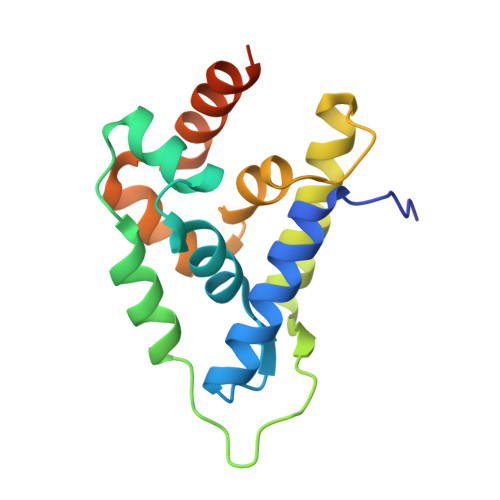
Atomic Structure of the Leishmania spp. Hsp100 N-Domain


Experimental Data Snapshot

  • Method: X-RAY DIFFRACTION
  • Resolution: 1.05 Å
  • R-Value Free: 0.153 
  • R-Value Work: 0.134 
  • R-Value Observed: 0.135 

wwPDB Validation   3D Report Full Report


This is version 1.2 of the entry. See complete history


Literature

Atomic structure of the Leishmania spp. Hsp100 N-domain.

Mercado, J.M.Lee, S.Chang, C.Sung, N.Soong, L.Catic, A.Tsai, F.T.F.

(2022) Proteins 90: 1242-1246

  • DOI: https://doi.org/10.1002/prot.26310
  • Primary Citation of Related Structures:  
    7TFM

  • PubMed Abstract: 

    Hsp100 is an ATP-dependent unfoldase that promotes protein disaggregation or facilitates the unfolding of aggregation-prone polypeptides marked for degradation. Recently, new Hsp100 functions are emerging. In Plasmodium, an Hsp100 drives malaria protein export, presenting a novel drug target. Whether Hsp100 has a similar function in other protists is unknown. We present the 1.06 Å resolution crystal structure of the Hsp100 N-domain from Leishmania spp., the causative agent of leishmaniasis in humans. Our structure reveals a network of methionines and aromatic amino acids that define the putative substrate-binding site and likely evolved to protect Hsp100 from oxidative damage in host immune cells.


  • Organizational Affiliation

    Department of Molecular and Cellular Biology, Baylor College of Medicine, Houston, Texas, USA.


Macromolecules
Find similar proteins by:  (by identity cutoff)  |  3D Structure
Entity ID: 1
MoleculeChains Sequence LengthOrganismDetailsImage
ATP-dependent Clp protease subunit, heat shock protein 100 (HSP100)158Leishmania mexicana MHOM/GT/2001/U1103Mutation(s): 0 
Gene Names: LMXM_08_29_1270
UniProt
Find proteins for E9ALU6 (Leishmania mexicana (strain MHOM/GT/2001/U1103))
Explore E9ALU6 
Go to UniProtKB:  E9ALU6
Entity Groups  
Sequence Clusters30% Identity50% Identity70% Identity90% Identity95% Identity100% Identity
UniProt GroupE9ALU6
Sequence Annotations
Expand
  • Reference Sequence
Small Molecules
Ligands 1 Unique
IDChains Name / Formula / InChI Key2D Diagram3D Interactions
GOL
Query on GOL

Download Ideal Coordinates CCD File 
B [auth A]GLYCEROL
C3 H8 O3
PEDCQBHIVMGVHV-UHFFFAOYSA-N
Experimental Data & Validation

Experimental Data

  • Method: X-RAY DIFFRACTION
  • Resolution: 1.05 Å
  • R-Value Free: 0.153 
  • R-Value Work: 0.134 
  • R-Value Observed: 0.135 
  • Space Group: P 41 21 2
Unit Cell:
Length ( Å )Angle ( ˚ )
a = 60.971α = 90
b = 60.971β = 90
c = 71.807γ = 90
Software Package:
Software NamePurpose
PHENIXrefinement
PDB_EXTRACTdata extraction
HKL-3000data reduction
HKL-3000data scaling
PHASERphasing

Structure Validation

View Full Validation Report


Entry History & Funding Information

Deposition Data


Funding OrganizationLocationGrant Number
National Institutes of Health/National Institute of General Medical Sciences (NIH/NIGMS)United StatesR01-GM104980
National Institutes of Health/National Institute of General Medical Sciences (NIH/NIGMS)United StatesR01-GM142143
National Institutes of Health/National Institute of Diabetes and Digestive and Kidney Disease (NIH/NIDDK)United StatesR01-DK115454
Cancer Prevention and Research Institute of Texas (CPRIT)United StatesRR140038
National Institutes of Health/National Institute of General Medical Sciences (NIH/NIGMS)United StatesS10-OD030246
Welch FoundationUnited StatesQ-1530-20190330
Department of Energy (DOE, United States)United StatesDE-AC02-06CH11357
National Institutes of Health/National Institute of General Medical Sciences (NIH/NIGMS)United StatesT32-GM008289

Revision History  (Full details and data files)

  • Version 1.0: 2022-02-16
    Type: Initial release
  • Version 1.1: 2022-04-27
    Changes: Database references
  • Version 1.2: 2023-10-18
    Changes: Data collection, Refinement description