Characterizing inhibitors of human AP endonuclease 1.
Pidugu, L.S., Servius, H.W., Sevdalis, S.E., Cook, M.E., Varney, K.M., Pozharski, E., Drohat, A.C.(2023) PLoS One 18: e0280526-e0280526
- PubMed: 36652434 Search on PubMedSearch on PubMed Central
- DOI: https://doi.org/10.1371/journal.pone.0280526
- Primary Citation Related Structures:
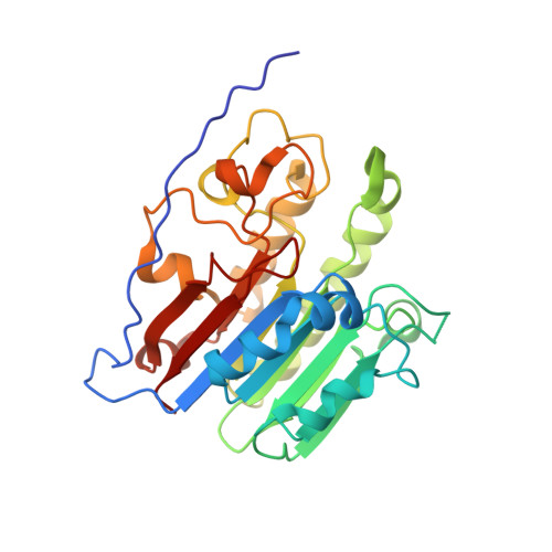
7TC2, 7TC3 - PubMed Abstract:
AP endonuclease 1 (APE1) processes DNA lesions including apurinic/apyrimidinic sites and 3´-blocking groups, mediating base excision repair and single strand break repair. Much effort has focused on developing specific inhibitors of APE1, which could have important applications in basic research and potentially lead to clinical anticancer agents. We used structural, biophysical, and biochemical methods to characterize several reported inhibitors, including 7-nitroindole-2-carboxylic acid (CRT0044876), given its small size, reported potency, and widespread use for studying APE1. Intriguingly, NMR chemical shift perturbation (CSP) experiments show that CRT0044876 and three similar indole-2-carboxylic acids bind a pocket distal from the APE1 active site. A crystal structure confirms these findings and defines the pose for 5-nitroindole-2-carboxylic acid. However, dynamic light scattering experiments show the indole compounds form colloidal aggregates that could bind (sequester) APE1, causing nonspecific inhibition. Endonuclease assays show the compounds lack significant APE1 inhibition under conditions (detergent) that disrupt aggregation. Thus, binding of the indole-2-carboxylic acids at the remote pocket does not inhibit APE1 repair activity. Myricetin also forms aggregates and lacks APE1 inhibition under aggregate-disrupting conditions. Two other reported compounds (MLS000552981, MLS000419194) inhibit APE1 in vitro with low micromolar IC50 and do not appear to aggregate in this concentration range. However, NMR CSP experiments indicate the compounds do not bind specifically to apo- or Mg2+-bound APE1, pointing to a non-specific mode of inhibition, possibly DNA binding. Our results highlight methods for rigorous interrogation of putative APE1 inhibitors and should facilitate future efforts to discover compounds that specifically inhibit this important repair enzyme.
- Department of Biochemistry and Molecular Biology, University of Maryland School of Medicine, Baltimore, Maryland, United States of America.
Organizational Affiliation: