Fluorescence-Based Activity Screening Assay Reveals Small Molecule Inhibitors of Vaccinia Virus mRNA Decapping Enzyme D9.
Bednarczyk, M., Peters, J.K., Kasprzyk, R., Starek, J., Warminski, M., Spiewla, T., Mugridge, J.S., Gross, J.D., Jemielity, J., Kowalska, J.(2022) ACS Chem Biol 17: 1460-1471
- PubMed: 35576528 Search on PubMedSearch on PubMed Central
- DOI: https://doi.org/10.1021/acschembio.2c00049
- Primary Citation Related Structures:
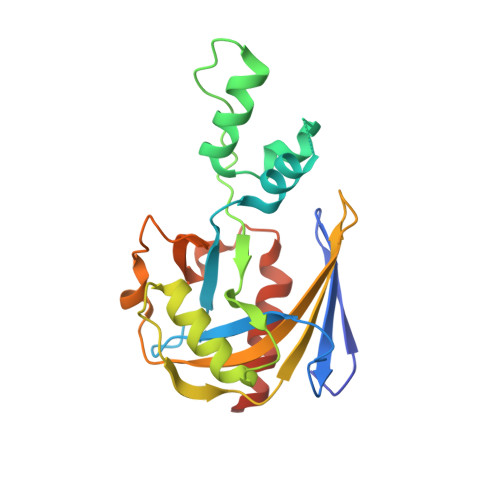
7T7H - PubMed Abstract:
Vaccinia virus (VACV) represents a family of poxviruses, which possess their own decapping machinery as a part of their strategy to eliminate host mRNAs and evade the innate immune response. D9 is one of the two encoded VACV decapping enzymes that is responsible for cap removal from the 5' end of both host mRNA transcripts and viral double-stranded RNAs. Little is known about the structural requirements for D9 inhibition by small molecules. Here, we identified a minimal D9 substrate and used it to develop a real-time fluorescence assay for inhibitor discovery and characterization. We screened a panel of nucleotide-derived substrate analogues and pharmacologically active candidates to identify several compounds with nano- and low micromolar IC 50 values. m 7 GpppCH 2 p was the most potent nucleotide inhibitor (IC 50 ∼ 0.08 μM), and seliciclib and CP-100356 were the most potent drug-like compounds (IC 50 0.57 and 2.7 μM, respectively). The hits identified through screening inhibited D9-catalyzed decapping of 26 nt RNA substrates but were not active toward VACV D10 or human decapping enzyme, Dcp1/2. The inhibition mode for one of the compounds (CP-100356) was elucidated based on the X-ray cocrystal structure, opening the possibility for structure-based design of novel D9 inhibitors and binding probes.
- Division of Biophysics, Institute of Experimental Physics, Faculty of Physics, University of Warsaw, Pasteura 5, Warsaw 02-093, Poland.
Organizational Affiliation: