7T5D

Neutron structure of Neurospora crassa Lytic Polysaccharide Monooxygenase 9D (NcLPMO9D) ascorbate soak

  • Classification: OXIDOREDUCTASE
  • Organism(s): Neurospora crassa
  • Expression System: Komagataella pastoris
  • Mutation(s): No 

  • Deposited: 2021-12-11 Released: 2022-12-28 
  • Deposition Author(s): Schroder, G.C., Meilleur, F.
  • Funding Organization(s): National Institute of Food and Agriculture (NIFA, United States), National Research Foundation in South Africa, Department of Energy (DOE, United States)

Experimental Data Snapshot

  • Method: NEUTRON DIFFRACTION
  • Resolution: 2.40 Å
  • R-Value Free: 0.309 
  • R-Value Work: 0.229 
  • R-Value Observed: 0.233 

wwPDB Validation   3D Report Full Report


This is version 1.1 of the entry. See complete history


Literature

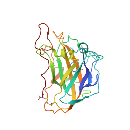
Capture of activated dioxygen intermediates at the copper-active site of a lytic polysaccharide monooxygenase.

Schroder, G.C.O'Dell, W.B.Webb, S.P.Agarwal, P.K.Meilleur, F.

(2022) Chem Sci 13: 13303-13320

  • DOI: https://doi.org/10.1039/d2sc05031e
  • Primary Citation of Related Structures:  
    7T5C, 7T5D, 7T5E

  • PubMed Abstract: 

    Metalloproteins perform a diverse array of redox-related reactions facilitated by the increased chemical functionality afforded by their metallocofactors. Lytic polysaccharide monooxygenases (LPMOs) are a class of copper-dependent enzymes that are responsible for the breakdown of recalcitrant polysaccharides via oxidative cleavage at the glycosidic bond. The activated copper-oxygen intermediates and their mechanism of formation remains to be established. Neutron protein crystallography which permits direct visualization of protonation states was used to investigate the initial steps of oxygen activation directly following active site copper reduction in Neurospora crassa LPMO9D. Herein, we cryo-trap an activated dioxygen intermediate in a mixture of superoxo and hydroperoxo states, and we identify the conserved second coordination shell residue His157 as the proton donor. Density functional theory calculations indicate that both superoxo and hydroperoxo active site states are stable. The hydroperoxo formed is potentially an early LPMO catalytic reaction intermediate or the first step in the mechanism of hydrogen peroxide formation in the absence of substrate. We observe that the N-terminal amino group of the copper coordinating His1 remains doubly protonated directly following molecular oxygen reduction by copper. Aided by molecular dynamics and mining minima free energy calculations we establish that the conserved second-shell His161 in Mt PMO3* displays conformational flexibility in solution and that this flexibility is also observed, though to a lesser extent, in His157 of Nc LPMO9D. The imidazolate form of His157 observed in our structure following oxygen intermediate protonation can be attributed to abolished His157 flexibility due steric hindrance in the crystal as well as the solvent-occluded active site environment due to crystal packing. A neutron crystal structure of Nc LPMO9D at low pH further supports occlusion of the active site since His157 remains singly protonated even at acidic conditions.


  • Organizational Affiliation

    Department of Molecular and Structural Biochemistry, North Carolina State University Raleigh NC 27695 USA fmeille@ncsu.edu.


Macromolecules
Find similar proteins by:  (by identity cutoff)  |  3D Structure
Entity ID: 1
MoleculeChains Sequence LengthOrganismDetailsImage
Lytic polysaccharide monooxygenase
A, B
223Neurospora crassaMutation(s): 0 
Gene Names: G15G9.090GE21DRAFT_7469
UniProt
Find proteins for Q1K8B6 (Neurospora crassa (strain ATCC 24698 / 74-OR23-1A / CBS 708.71 / DSM 1257 / FGSC 987))
Explore Q1K8B6 
Go to UniProtKB:  Q1K8B6
Entity Groups  
Sequence Clusters30% Identity50% Identity70% Identity90% Identity95% Identity100% Identity
UniProt GroupQ1K8B6
Sequence Annotations
Expand
  • Reference Sequence
Oligosaccharides

Help

Entity ID: 2
MoleculeChains Length2D Diagram Glycosylation3D Interactions
2-acetamido-2-deoxy-beta-D-glucopyranose-(1-4)-2-acetamido-2-deoxy-beta-D-glucopyranose
C, D
2N-Glycosylation
Glycosylation Resources
GlyTouCan:  G42666HT
GlyCosmos:  G42666HT
GlyGen:  G42666HT
Experimental Data & Validation

Experimental Data

  • Method: NEUTRON DIFFRACTION
  • Resolution: 2.40 Å
  • R-Value Free: 0.309 
  • R-Value Work: 0.229 
  • R-Value Observed: 0.233 
  • Space Group: P 1 21 1
Unit Cell:
Length ( Å )Angle ( ˚ )
a = 67.73α = 90
b = 42.18β = 99
c = 69.76γ = 90
Software Package:
Software NamePurpose
PHENIXrefinement
Mantiddata reduction
LaueViewdata scaling
PHASERphasing
Cootmodel building

Structure Validation

View Full Validation Report


Entry History & Funding Information

Deposition Data


Funding OrganizationLocationGrant Number
National Institute of Food and Agriculture (NIFA, United States)South AfricaHatch 211001
National Research Foundation in South AfricaSouth Africa--
Department of Energy (DOE, United States)South Africa--

Revision History  (Full details and data files)

  • Version 1.0: 2022-12-28
    Type: Initial release
  • Version 1.1: 2023-10-25
    Changes: Data collection, Refinement description