7SZ3

Mouse PARP13/ZAP ZnF5-WWE1-WWE2 bound to ADPr


Experimental Data Snapshot

  • Method: X-RAY DIFFRACTION
  • Resolution: 2.20 Å
  • R-Value Free: 0.231 
  • R-Value Work: 0.193 
  • R-Value Observed: 0.196 

wwPDB Validation   3D Report Full Report


Ligand Structure Quality Assessment 


This is version 1.1 of the entry. See complete history


Literature

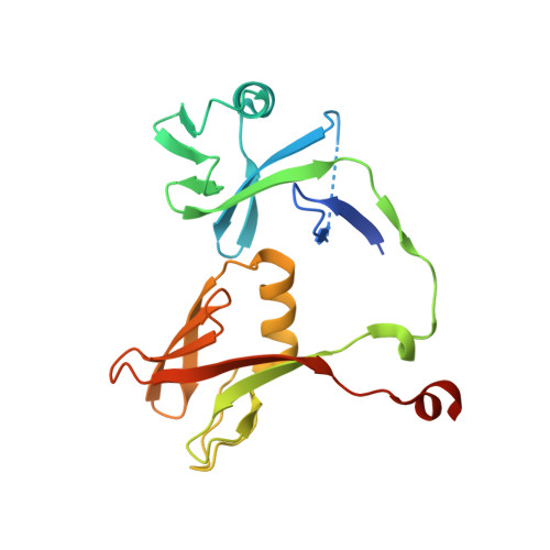
Crystal structures and functional analysis of the ZnF5-WWE1-WWE2 region of PARP13/ZAP define a distinctive mode of engaging poly(ADP-ribose).

Kuttiyatveetil, J.R.A.Soufari, H.Dasovich, M.Uribe, I.R.Mirhasan, M.Cheng, S.J.Leung, A.K.L.Pascal, J.M.

(2022) Cell Rep 41: 111529-111529

  • DOI: https://doi.org/10.1016/j.celrep.2022.111529
  • Primary Citation of Related Structures:  
    7SZ2, 7SZ3

  • PubMed Abstract: 

    PARP13/ZAP (zinc-finger antiviral protein) acts against multiple viruses by promoting degradation of viral mRNA. PARP13 has four N-terminal zinc (Zn) fingers that bind CG-rich nucleotide sequences, a C-terminal ADP ribosyltransferase fold, and a central region with a fifth Zn finger and tandem WWE domains. The central PARP13 region, ZnF5-WWE1-WWE2, is implicated in binding poly(ADP-ribose); however, there are limited insights into its structure and function. We present crystal structures of ZnF5-WWE1-WWE2 from mouse PARP13 in complex with ADP-ribose and in complex with ATP. The crystal structures and binding studies demonstrate that WWE2 interacts with ADP-ribose and ATP, whereas WWE1 does not have a functional binding site. Binding studies with poly(ADP-ribose) ligands indicate that WWE2 serves as an anchor for preferential binding to the terminal end of poly(ADP-ribose) chains. The composite ZnF5-WWE1-WWE2 structure forms an extended surface to engage ADP-ribose chains, representing a distinctive mode of recognition that provides a framework for investigating the impact of poly(ADP-ribose) on PARP13 function.


  • Organizational Affiliation

    Department of Biochemistry and Molecular Medicine, Université de Montréal, Montréal, QC H3T 1J4, Canada.


Macromolecules
Find similar proteins by:  (by identity cutoff)  |  3D Structure
Entity ID: 1
MoleculeChains Sequence LengthOrganismDetailsImage
Zinc finger CCCH-type antiviral protein 1
A, B
198Mus musculusMutation(s): 0 
Gene Names: Zc3hav1
UniProt
Find proteins for Q3UPF5 (Mus musculus)
Explore Q3UPF5 
Go to UniProtKB:  Q3UPF5
Entity Groups  
Sequence Clusters30% Identity50% Identity70% Identity90% Identity95% Identity100% Identity
UniProt GroupQ3UPF5
Sequence Annotations
Expand
  • Reference Sequence
Small Molecules
Ligands 3 Unique
IDChains Name / Formula / InChI Key2D Diagram3D Interactions
APR (Subject of Investigation/LOI)
Query on APR

Download Ideal Coordinates CCD File 
D [auth A],
I [auth B]
ADENOSINE-5-DIPHOSPHORIBOSE
C15 H23 N5 O14 P2
SRNWOUGRCWSEMX-KEOHHSTQSA-N
PO4
Query on PO4

Download Ideal Coordinates CCD File 
F [auth A]
G [auth A]
H [auth A]
J [auth B]
K [auth B]
F [auth A],
G [auth A],
H [auth A],
J [auth B],
K [auth B],
L [auth B],
M [auth B]
PHOSPHATE ION
O4 P
NBIIXXVUZAFLBC-UHFFFAOYSA-K
GOL
Query on GOL

Download Ideal Coordinates CCD File 
C [auth A],
E [auth A]
GLYCEROL
C3 H8 O3
PEDCQBHIVMGVHV-UHFFFAOYSA-N
Experimental Data & Validation

Experimental Data

  • Method: X-RAY DIFFRACTION
  • Resolution: 2.20 Å
  • R-Value Free: 0.231 
  • R-Value Work: 0.193 
  • R-Value Observed: 0.196 
  • Space Group: P 31 2 1
Unit Cell:
Length ( Å )Angle ( ˚ )
a = 87.415α = 90
b = 87.415β = 90
c = 128.741γ = 120
Software Package:
Software NamePurpose
Aimlessdata scaling
REFMACrefinement
PDB_EXTRACTdata extraction
XDSdata reduction
Cootmodel building
PHASERphasing

Structure Validation

View Full Validation Report


Ligand Structure Quality Assessment 


Entry History & Funding Information

Deposition Data


Funding OrganizationLocationGrant Number
Canadian Institutes of Health Research (CIHR)Canada--

Revision History  (Full details and data files)

  • Version 1.0: 2022-10-05
    Type: Initial release
  • Version 1.1: 2022-11-09
    Changes: Database references