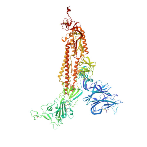
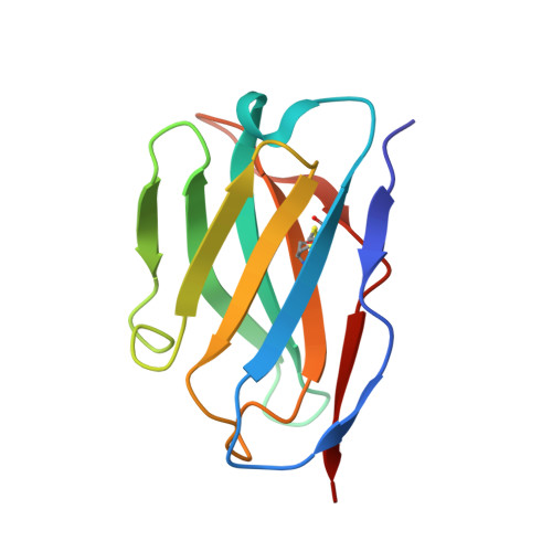
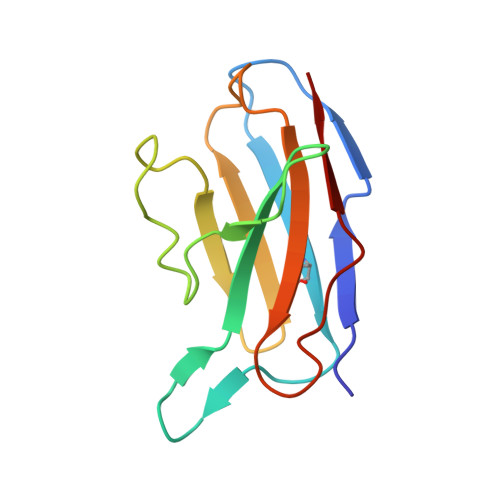
7SWX

SARS-CoV-2 Spike in complex with neutralizing Fab SARS2-57 (three down conformation)


Experimental Data Snapshot

  • Method: ELECTRON MICROSCOPY
  • Resolution: 3.13 Å
  • Aggregation State: PARTICLE 
  • Reconstruction Method: SINGLE PARTICLE 

wwPDB Validation   3D Report Full Report


This is version 1.1 of the entry. See complete history


Literature

A protective antibody targeting the N-terminal domain of SARS-CoV-2 spike recognizes most emerging variants

Adams, L.J.VanBlargan, L.A.Liu, Z.Gilchuk, P.Zhao, H.Chen, R.E.Raju, S.Whitener, B.Shrihari, S.Jethva, P.N.Gross, M.L.Crowe, J.E.Whelan, S.P.J.Diamond, M.S.Fremont, D.H.

To be published.

Macromolecules
Find similar proteins by:  (by identity cutoff)  |  3D Structure
Entity ID: 1
MoleculeChains Sequence LengthOrganismDetailsImage
Spike glycoprotein
A, B, C
1,133Severe acute respiratory syndrome coronavirus 2Mutation(s): 6 
Gene Names: S2
UniProt
Find proteins for P0DTC2 (Severe acute respiratory syndrome coronavirus 2)
Explore P0DTC2 
Go to UniProtKB:  P0DTC2
Entity Groups  
Sequence Clusters30% Identity50% Identity70% Identity90% Identity95% Identity100% Identity
UniProt GroupP0DTC2
Sequence Annotations
Expand
  • Reference Sequence
Find similar proteins by:  (by identity cutoff)  |  3D Structure
Entity ID: 2
MoleculeChains Sequence LengthOrganismDetailsImage
SARS2-57 Fv heavy chainD [auth H]117Mus musculusMutation(s): 0 
Entity Groups  
Sequence Clusters30% Identity50% Identity70% Identity90% Identity95% Identity100% Identity
Sequence Annotations
Expand
  • Reference Sequence
Find similar proteins by:  (by identity cutoff)  |  3D Structure
Entity ID: 3
MoleculeChains Sequence LengthOrganismDetailsImage
SARS2-57 Fv light chainE [auth L]112Mus musculusMutation(s): 0 
Entity Groups  
Sequence Clusters30% Identity50% Identity70% Identity90% Identity95% Identity100% Identity
Sequence Annotations
Expand
  • Reference Sequence
Oligosaccharides

Help

Entity ID: 4
MoleculeChains Length2D Diagram Glycosylation3D Interactions
2-acetamido-2-deoxy-beta-D-glucopyranose-(1-4)-2-acetamido-2-deoxy-beta-D-glucopyranose
AA [auth a],
BA [auth b],
CA [auth c],
DA [auth d],
EA [auth e],
AA [auth a],
BA [auth b],
CA [auth c],
DA [auth d],
EA [auth e],
F [auth D],
FA [auth f],
G [auth E],
GA [auth g],
H [auth F],
I [auth G],
J [auth I],
K [auth J],
L [auth K],
M,
N,
O,
P,
Q,
R,
S,
T,
U,
V,
W,
X,
Y,
Z
2N/AN-Glycosylation
Glycosylation Resources
GlyTouCan:  G42666HT
GlyCosmos:  G42666HT
GlyGen:  G42666HT
Small Molecules
Ligands 1 Unique
IDChains Name / Formula / InChI Key2D Diagram3D Interactions
NAG
Query on NAG

Download Ideal Coordinates CCD File 
AB [auth C]
BB [auth C]
CB [auth C]
DB [auth C]
EB [auth C]
AB [auth C],
BB [auth C],
CB [auth C],
DB [auth C],
EB [auth C],
FB [auth C],
HA [auth A],
IA [auth A],
JA [auth A],
KA [auth A],
LA [auth A],
MA [auth A],
NA [auth A],
OA [auth B],
PA [auth B],
QA [auth B],
RA [auth B],
SA [auth B],
TA [auth B],
UA [auth B],
VA [auth B],
WA [auth B],
XA [auth C],
YA [auth C],
ZA [auth C]
2-acetamido-2-deoxy-beta-D-glucopyranose
C8 H15 N O6
OVRNDRQMDRJTHS-FMDGEEDCSA-N
Experimental Data & Validation

Experimental Data

  • Method: ELECTRON MICROSCOPY
  • Resolution: 3.13 Å
  • Aggregation State: PARTICLE 
  • Reconstruction Method: SINGLE PARTICLE 

Structure Validation

View Full Validation Report


Entry History & Funding Information

Deposition Data


Funding OrganizationLocationGrant Number
National Institutes of Health/National Institute Of Allergy and Infectious Diseases (NIH/NIAID)United States75N93019C00062
National Institutes of Health/National Institute Of Allergy and Infectious Diseases (NIH/NIAID)United StatesHHSN272201700060C

Revision History  (Full details and data files)

  • Version 1.0: 2022-11-23
    Type: Initial release
  • Version 1.1: 2023-02-01
    Changes: Other, Structure summary