7SWS

Crystal structure of the chromoprotein amilCP


Experimental Data Snapshot

  • Method: X-RAY DIFFRACTION
  • Resolution: 1.64 Å
  • R-Value Free: 0.218 
  • R-Value Work: 0.194 
  • R-Value Observed: 0.195 

wwPDB Validation   3D Report Full Report


This is version 2.0 of the entry. See complete history


Literature

Over the rainbow: structural characterization of the chromoproteins gfasPurple, amilCP, spisPink and eforRed.

Ahmed, F.H.Caputo, A.T.French, N.G.Peat, T.S.Whitfield, J.Warden, A.C.Newman, J.Scott, C.

(2022) Acta Crystallogr D Struct Biol 78: 599-612

  • DOI: https://doi.org/10.1107/S2059798322002625
  • Primary Citation of Related Structures:  
    7SWR, 7SWS, 7SWT, 7SWU

  • PubMed Abstract: 

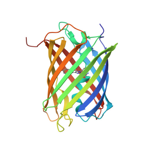
    Anthozoan chromoproteins are highly pigmented, diversely coloured and readily produced in recombinant expression systems. While they are a versatile and powerful building block in synthetic biology for applications such as biosensor development, they are not widely used in comparison to the related fluorescent proteins, partly due to a lack of structural characterization to aid protein engineering. Here, high-resolution X-ray crystal structures of four open-source chromoproteins, gfasPurple, amilCP, spisPink and eforRed, are presented. These proteins are dimers in solution, and mutation at the conserved dimer interface leads to loss of visible colour development in gfasPurple. The chromophores are trans and noncoplanar in gfasPurple, amilCP and spisPink, while that in eforRed is cis and noncoplanar, and also emits fluorescence. Like other characterized chromoproteins, gfasPurple, amilCP and eforRed contain an sp 2 -hybridized N-acylimine in the peptide bond preceding the chromophore, while spisPink is unusual and demonstrates a true sp 3 -hybridized trans-peptide bond at this position. It was found that point mutations at the chromophore-binding site in gfasPurple that substitute similar amino acids to those in amilCP and spisPink generate similar colours. These features and observations have implications for the utility of these chromoproteins in protein engineering and synthetic biology applications.


  • Organizational Affiliation

    Land and Water, CSIRO, Clunies Ross Street, Canberra, ACT 2601, Australia.


Macromolecules
Find similar proteins by:  (by identity cutoff)  |  3D Structure
Entity ID: 1
MoleculeChains Sequence LengthOrganismDetailsImage
Chromoprotein amilCP
A, B, C, D, E
A, B, C, D, E, F, G, H
219Acropora milleporaMutation(s): 0 
UniProt
Find proteins for Q66PV0 (Acropora millepora)
Explore Q66PV0 
Go to UniProtKB:  Q66PV0
Entity Groups  
Sequence Clusters30% Identity50% Identity70% Identity90% Identity95% Identity100% Identity
UniProt GroupQ66PV0
Sequence Annotations
Expand
  • Reference Sequence
Small Molecules
Ligands 2 Unique
IDChains Name / Formula / InChI Key2D Diagram3D Interactions
BR
Query on BR

Download Ideal Coordinates CCD File 
L [auth C]BROMIDE ION
Br
CPELXLSAUQHCOX-UHFFFAOYSA-M
CL
Query on CL

Download Ideal Coordinates CCD File 
I [auth A]
J [auth B]
K [auth C]
M [auth D]
N [auth E]
I [auth A],
J [auth B],
K [auth C],
M [auth D],
N [auth E],
O [auth F],
P [auth G],
Q [auth H],
R [auth H]
CHLORIDE ION
Cl
VEXZGXHMUGYJMC-UHFFFAOYSA-M
Modified Residues  1 Unique
IDChains TypeFormula2D DiagramParent
CRQ
Query on CRQ
A, B, C, D, E
A, B, C, D, E, F, G, H
L-PEPTIDE LINKINGC16 H16 N4 O5GLN, TYR, GLY
Experimental Data & Validation

Experimental Data

  • Method: X-RAY DIFFRACTION
  • Resolution: 1.64 Å
  • R-Value Free: 0.218 
  • R-Value Work: 0.194 
  • R-Value Observed: 0.195 
  • Space Group: P 1 21 1
Unit Cell:
Length ( Å )Angle ( ˚ )
a = 71.524α = 90
b = 131.742β = 100.75
c = 93.671γ = 90
Software Package:
Software NamePurpose
BUSTERrefinement
XDSdata reduction
Aimlessdata scaling
PHASERphasing

Structure Validation

View Full Validation Report


Entry History & Funding Information

Deposition Data


Funding OrganizationLocationGrant Number
Other government--

Revision History  (Full details and data files)

  • Version 1.0: 2022-04-20
    Type: Initial release
  • Version 1.1: 2022-05-18
    Changes: Database references
  • Version 1.2: 2023-10-18
    Changes: Data collection, Refinement description
  • Version 2.0: 2023-11-15
    Changes: Atomic model, Data collection, Derived calculations