7SMC

p107 pocket domain complexed with ARID4A peptide


Experimental Data Snapshot

  • Method: X-RAY DIFFRACTION
  • Resolution: 2.70 Å
  • R-Value Free: 0.298 
  • R-Value Work: 0.238 
  • R-Value Observed: 0.241 

wwPDB Validation   3D Report Full Report


This is version 1.2 of the entry. See complete history


Literature

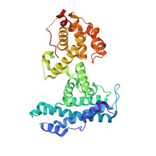
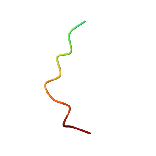
Structural basis for tunable affinity and specificity of LxCxE-dependent protein interactions with the retinoblastoma protein family.

Putta, S.Alvarez, L.Ludtke, S.Sehr, P.Muller, G.A.Fernandez, S.M.Tripathi, S.Lewis, J.Gibson, T.J.Chemes, L.B.Rubin, S.M.

(2022) Structure 30: 1340

  • DOI: https://doi.org/10.1016/j.str.2022.05.019
  • Primary Citation of Related Structures:  
    7SMC, 7SMD, 7SME, 7SMF

  • PubMed Abstract: 

    The retinoblastoma protein (Rb) and its homologs p107 and p130 are critical regulators of gene expression during the cell cycle and are commonly inactivated in cancer. Rb proteins use their "pocket domain" to bind an LxCxE sequence motif in other proteins, many of which function with Rb proteins to co-regulate transcription. Here, we present binding data and crystal structures of the p107 pocket domain in complex with LxCxE peptides from the transcriptional co-repressor proteins HDAC1, ARID4A, and EID1. Our results explain why Rb and p107 have weaker affinity for cellular LxCxE proteins compared with the E7 protein from human papillomavirus, which has been used as the primary model for understanding LxCxE motif interactions. Our structural and mutagenesis data also identify and explain differences in Rb and p107 affinities for some LxCxE-containing sequences. Our study provides new insights into how Rb proteins bind their cell partners with varying affinity and specificity.


  • Organizational Affiliation

    Department of Chemistry and Biochemistry, University of California, Santa Cruz, CA 95064, USA.


Macromolecules
Find similar proteins by:  (by identity cutoff)  |  3D Structure
Entity ID: 1
MoleculeChains Sequence LengthOrganismDetailsImage
Retinoblastoma-like protein 1
A, C
371Homo sapiensMutation(s): 0 
Gene Names: RBL1
UniProt & NIH Common Fund Data Resources
Find proteins for P28749 (Homo sapiens)
Explore P28749 
Go to UniProtKB:  P28749
PHAROS:  P28749
GTEx:  ENSG00000080839 
Entity Groups  
Sequence Clusters30% Identity50% Identity70% Identity90% Identity95% Identity100% Identity
UniProt GroupP28749
Sequence Annotations
Expand
  • Reference Sequence

Find similar proteins by:  Sequence   |   3D Structure  

Entity ID: 2
MoleculeChains Sequence LengthOrganismDetailsImage
AT-rich interactive domain-containing protein 4A
B, D
15Homo sapiensMutation(s): 0 
UniProt & NIH Common Fund Data Resources
Find proteins for P29374 (Homo sapiens)
Explore P29374 
Go to UniProtKB:  P29374
PHAROS:  P29374
GTEx:  ENSG00000032219 
Entity Groups  
Sequence Clusters30% Identity50% Identity70% Identity90% Identity95% Identity100% Identity
UniProt GroupP29374
Sequence Annotations
Expand
  • Reference Sequence
Experimental Data & Validation

Experimental Data

  • Method: X-RAY DIFFRACTION
  • Resolution: 2.70 Å
  • R-Value Free: 0.298 
  • R-Value Work: 0.238 
  • R-Value Observed: 0.241 
  • Space Group: P 1
Unit Cell:
Length ( Å )Angle ( ˚ )
a = 62.863α = 69.89
b = 62.923β = 76
c = 71.274γ = 74.36
Software Package:
Software NamePurpose
PHENIXrefinement
XDSdata reduction
Aimlessdata scaling
PHASERphasing

Structure Validation

View Full Validation Report


Entry History & Funding Information

Deposition Data


Funding OrganizationLocationGrant Number
National Institutes of Health/National Institute of General Medical Sciences (NIH/NIGMS)United StatesGM124148

Revision History  (Full details and data files)

  • Version 1.0: 2022-06-29
    Type: Initial release
  • Version 1.1: 2022-09-14
    Changes: Database references
  • Version 1.2: 2023-10-18
    Changes: Data collection, Refinement description