7SHQ | pdb_00007shq

Structure of a functional construct of eukaryotic elongation factor 2 kinase in complex with calmodulin.


Experimental Data Snapshot

  • Method: X-RAY DIFFRACTION
  • Resolution: 2.34 Å
  • R-Value Free: 
    0.252 (Depositor), 0.265 (DCC) 
  • R-Value Work: 
    0.229 (Depositor) 
  • R-Value Observed: 
    0.230 (Depositor) 

Starting Models: experimental
View more details

wwPDB Validation 3D Report Full Report

Validation slider image for 7SHQ

This is version 1.3 of the entry. See complete history

Literature

Structural basis for the calmodulin-mediated activation of eukaryotic elongation factor 2 kinase.

Piserchio, A.Isiorho, E.A.Long, K.Bohanon, A.L.Kumar, E.A.Will, N.Jeruzalmi, D.Dalby, K.N.Ghose, R.

(2022) Sci Adv 8: eabo2039-eabo2039

  • DOI: https://doi.org/10.1126/sciadv.abo2039
  • Primary Citation Related Structures: 
    7SHQ

  • PubMed Abstract: 

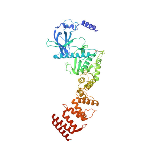
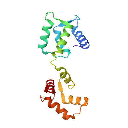
    Translation is a tightly regulated process that ensures optimal protein quality and enables adaptation to energy/nutrient availability. The α-kinase eukaryotic elongation factor 2 kinase (eEF-2K), a key regulator of translation, specifically phosphorylates the guanosine triphosphatase eEF-2, thereby reducing its affinity for the ribosome and suppressing the elongation phase of protein synthesis. eEF-2K activation requires calmodulin binding and autophosphorylation at the primary stimulatory site, T348. Biochemical studies predict a calmodulin-mediated activation mechanism for eEF-2K distinct from other calmodulin-dependent kinases. Here, we resolve the atomic details of this mechanism through a 2.3-Å crystal structure of the heterodimeric complex of calmodulin and the functional core of eEF-2K (eEF-2K TR ). This structure, which represents the activated T348-phosphorylated state of eEF-2K TR , highlights an intimate association of the kinase with the calmodulin C-lobe, creating an "activation spine" that connects its amino-terminal calmodulin-targeting motif to its active site through a conserved regulatory element.


  • Organizational Affiliation
    • Department of Chemistry and Biochemistry, The City College of New York, New York, NY 10031, USA.

Macromolecule Content 

  • Total Structure Weight: 77.37 kDa 
  • Atom Count: 5,268 
  • Modeled Residue Count: 637 
  • Deposited Residue Count: 679 
  • Unique protein chains: 2

Macromolecules

Find similar proteins by:|  3D Structure
Entity ID: 1
MoleculeChains  Sequence LengthOrganismDetailsImage
Eukaryotic elongation factor 2 kinase,Eukaryotic elongation factor 2 kinase531Homo sapiensMutation(s): 0 
Gene Names: EEF2K
EC: 2.7.11.20
UniProt & NIH Common Fund Data Resources
Find proteins for O00418 (Homo sapiens)
Explore O00418 
Go to UniProtKB:  O00418
PHAROS:  O00418
GTEx:  ENSG00000103319 
Entity Groups
Sequence Clusters30% Identity50% Identity70% Identity90% Identity95% Identity100% Identity
UniProt GroupO00418
Sequence Annotations
Expand
Reference Sequence
Find similar proteins by:|  3D Structure
Entity ID: 2
MoleculeChains  Sequence LengthOrganismDetailsImage
Calmodulin-1148Homo sapiensMutation(s): 0 
Gene Names: CALM1CALMCAMCAM1
UniProt & NIH Common Fund Data Resources
Find proteins for P0DP23 (Homo sapiens)
Explore P0DP23 
Go to UniProtKB:  P0DP23
PHAROS:  P0DP23
GTEx:  ENSG00000198668 
Entity Groups
Sequence Clusters30% Identity50% Identity70% Identity90% Identity95% Identity100% Identity
UniProt GroupP0DP23
Sequence Annotations
Expand
Reference Sequence

Small Molecules

Ligands 3 Unique
IDChains Name / Formula / InChI Key2D Diagram3D Interactions
ZN
(Subject of Investigation/LOI)

Query on ZN


Download:Ideal Coordinates CCD File
C [auth A]ZINC ION
Zn
PTFCDOFLOPIGGS-UHFFFAOYSA-N
CA
(Subject of Investigation/LOI)

Query on CA


Download:Ideal Coordinates CCD File
F [auth B],
G [auth B]
CALCIUM ION
Ca
BHPQYMZQTOCNFJ-UHFFFAOYSA-N
MG
(Subject of Investigation/LOI)

Query on MG


Download:Ideal Coordinates CCD File
D [auth A],
E [auth A],
H [auth B],
I [auth B],
J [auth B]
MAGNESIUM ION
Mg
JLVVSXFLKOJNIY-UHFFFAOYSA-N
Modified Residues  1 Unique
IDChains TypeFormula2D DiagramParent
TPO
Query on TPO
A
L-PEPTIDE LINKINGC4 H10 N O6 PTHR

Experimental Data & Validation

Experimental Data

  • Method: X-RAY DIFFRACTION
  • Resolution: 2.34 Å
  • R-Value Free:  0.252 (Depositor), 0.265 (DCC) 
  • R-Value Work:  0.229 (Depositor) 
  • R-Value Observed: 0.230 (Depositor) 
Space Group: P 31 2 1
Unit Cell:
Length ( Å )Angle ( ˚ )
a = 58.495α = 90
b = 58.495β = 90
c = 365.779γ = 120
Software Package:
Software NamePurpose
PHENIXrefinement
ISOLDErefinement
Cootmodel building
MR-Rosettaphasing
autoPROCdata scaling
autoPROCdata reduction

Structure Validation

View Full Validation Report


Entry History 

& Funding Information

Deposition Data


Funding OrganizationLocationGrant Number
National Institutes of Health/National Institute of General Medical Sciences (NIH/NIGMS)United StatesGM084278

Revision History  (Full details and data files)

  • Version 1.0: 2022-04-06
    Type: Initial release
  • Version 1.1: 2022-08-03
    Changes: Database references
  • Version 1.2: 2023-10-18
    Changes: Data collection, Database references, Refinement description
  • Version 1.3: 2024-11-06
    Changes: Structure summary