7SGM

Crystal structure of a Fab variant containing a fluorescent noncanonical amino acid with blocked excited state proton transfer and in complex with its antigen, CD40L


Experimental Data Snapshot

  • Method: X-RAY DIFFRACTION
  • Resolution: 2.00 Å
  • R-Value Free: 0.228 
  • R-Value Work: 0.193 
  • R-Value Observed: 0.195 

wwPDB Validation   3D Report Full Report


Ligand Structure Quality Assessment 


This is version 1.3 of the entry. See complete history


Literature

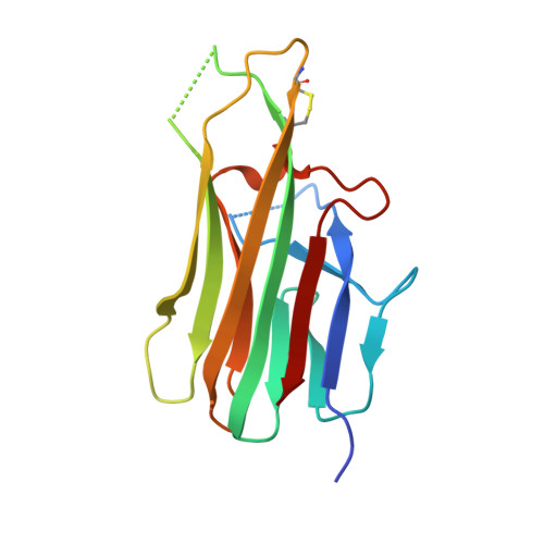
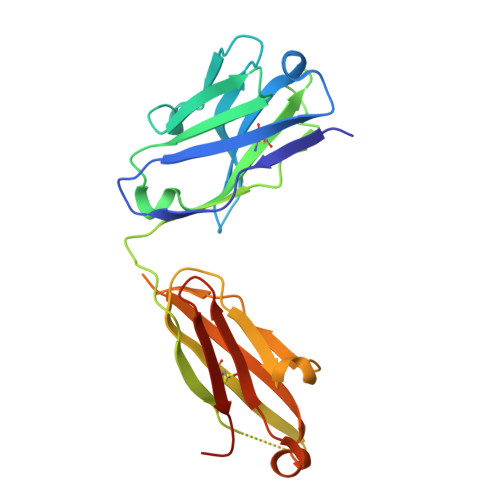
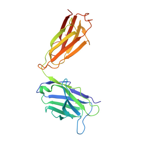
Structural Basis for Blocked Excited State Proton Transfer in a Fluorescent, Photoacidic Non-Canonical Amino Acid-Containing Antibody Fragment.

Henderson, J.N.Simmons, C.R.Mills, J.H.

(2022) J Mol Biol 434: 167455-167455

  • DOI: https://doi.org/10.1016/j.jmb.2022.167455
  • Primary Citation of Related Structures:  
    7SEN, 7SGM

  • PubMed Abstract: 

    The fluorescent non-canonical amino acid (fNCAA) L-(7-hydroxycoumarin-4-yl)ethylglycine (7-HCAA) contains a photoacidic 7-hydroxycoumarin (7-HC) side chain whose fluorescence properties can be tuned by its environment. In proteins, many alterations to 7-HCAA's fluorescence spectra have been reported including increases and decreases in intensity and red- and blue-shifted emission maxima. The ability to rationally design protein environments that alter 7-HCAA's fluorescence properties in predictable ways could lead to novel protein-based sensors of biological function. However, these efforts are likely limited by a lack of structural characterization of 7-HCAA-containing proteins. Here, we report the steady-state spectroscopic and x-ray crystallographic characterization of a 7-HCAA-containing antibody fragment (in the apo and antigen-bound forms) in which a substantially blue-shifted 7-HCAA emission maximum (∼70 nm) is observed relative to the free amino acid. Our structural characterization of these proteins provides evidence that the blue shift is a consequence of the fact that excited state proton transfer (ESPT) from the 7-HC phenol has been almost completely blocked by interactions with the protein backbone. Furthermore, a direct interaction between a residue in the antigen and the fluorophore served to further block proton transfer relative to the apoprotein. The structural basis of the unprecedented blue shift in 7-HCAA emission reported here provides a framework for the development of new fluorescent protein-based sensors.


  • Organizational Affiliation

    The Biodesign Center for Molecular Design and Biomimetics, Arizona State University, Tempe, AZ, USA.


Macromolecules
Find similar proteins by:  (by identity cutoff)  |  3D Structure
Entity ID: 1
MoleculeChains Sequence LengthOrganismDetailsImage
CD40 ligand
A, B, C
146Homo sapiensMutation(s): 0 
Gene Names: CD40LGCD40LTNFSF5TRAP
UniProt & NIH Common Fund Data Resources
Find proteins for P29965 (Homo sapiens)
Explore P29965 
Go to UniProtKB:  P29965
PHAROS:  P29965
GTEx:  ENSG00000102245 
Entity Groups  
Sequence Clusters30% Identity50% Identity70% Identity90% Identity95% Identity100% Identity
UniProt GroupP29965
Sequence Annotations
Expand
  • Reference Sequence
Find similar proteins by:  (by identity cutoff)  |  3D Structure
Entity ID: 2
MoleculeChains Sequence LengthOrganismDetailsImage
5c8* Fab heavy chainD [auth H],
F [auth K],
H [auth X]
226Homo sapiensMutation(s): 0 
Entity Groups  
Sequence Clusters30% Identity50% Identity70% Identity90% Identity95% Identity100% Identity
Sequence Annotations
Expand
  • Reference Sequence
Find similar proteins by:  (by identity cutoff)  |  3D Structure
Entity ID: 3
MoleculeChains Sequence LengthOrganismDetailsImage
5c8* Fab light chainE [auth L],
G [auth M],
I [auth Y]
218Homo sapiensMutation(s): 0 
Entity Groups  
Sequence Clusters30% Identity50% Identity70% Identity90% Identity95% Identity100% Identity
Sequence Annotations
Expand
  • Reference Sequence
Small Molecules
Ligands 4 Unique
IDChains Name / Formula / InChI Key2D Diagram3D Interactions
NAG
Query on NAG

Download Ideal Coordinates CCD File 
J [auth A],
M [auth B],
O [auth C]
2-acetamido-2-deoxy-beta-D-glucopyranose
C8 H15 N O6
OVRNDRQMDRJTHS-FMDGEEDCSA-N
TRS
Query on TRS

Download Ideal Coordinates CCD File 
K [auth A]2-AMINO-2-HYDROXYMETHYL-PROPANE-1,3-DIOL
C4 H12 N O3
LENZDBCJOHFCAS-UHFFFAOYSA-O
PEG
Query on PEG

Download Ideal Coordinates CCD File 
BA [auth Y]
CA [auth Y]
L [auth A]
N [auth B]
P [auth C]
BA [auth Y],
CA [auth Y],
L [auth A],
N [auth B],
P [auth C],
Q [auth H],
R [auth H],
T [auth L],
U [auth L],
V [auth L],
W [auth K],
Y [auth M],
Z [auth X]
DI(HYDROXYETHYL)ETHER
C4 H10 O3
MTHSVFCYNBDYFN-UHFFFAOYSA-N
TMO
Query on TMO

Download Ideal Coordinates CCD File 
AA [auth Y],
S [auth L],
X [auth M]
trimethylamine oxide
C3 H9 N O
UYPYRKYUKCHHIB-UHFFFAOYSA-N
Experimental Data & Validation

Experimental Data

  • Method: X-RAY DIFFRACTION
  • Resolution: 2.00 Å
  • R-Value Free: 0.228 
  • R-Value Work: 0.193 
  • R-Value Observed: 0.195 
  • Space Group: C 1 2 1
Unit Cell:
Length ( Å )Angle ( ˚ )
a = 226.847α = 90
b = 131.252β = 109.1
c = 97.296γ = 90
Software Package:
Software NamePurpose
REFMACrefinement
HKL-2000data scaling
PDB_EXTRACTdata extraction
HKL-2000data reduction
PHASERphasing

Structure Validation

View Full Validation Report


Ligand Structure Quality Assessment 


Entry History & Funding Information

Deposition Data


Funding OrganizationLocationGrant Number
National Institutes of Health/National Institute of General Medical Sciences (NIH/NIGMS)United StatesR01 GM136996

Revision History  (Full details and data files)

  • Version 1.0: 2022-02-02
    Type: Initial release
  • Version 1.1: 2022-04-27
    Changes: Database references
  • Version 1.2: 2023-10-18
    Changes: Data collection, Refinement description
  • Version 1.3: 2023-11-15
    Changes: Data collection, Derived calculations