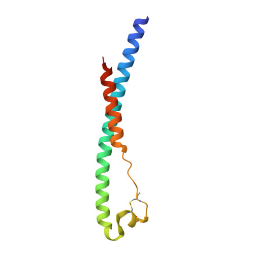
Structure of the Core Postfusion Porcine Endogenous Retrovirus Fusion Protein.
Dean, T.T., Serrao, V.H.B., Lee, J.E.(2022) mBio 13: e0292021-e0292021
- PubMed: 35073741 Search on PubMedSearch on PubMed Central
- DOI: https://doi.org/10.1128/mbio.02920-21
- Primary Citation Related Structures:
7S94 - PubMed Abstract:
Retroviral elements from endogenous retroviruses have functions in mammalian physiology. The best-known examples are the envelope proteins that function in placenta development and immune suppression. Porcine endogenous retroviruses (PERVs) are an understudied class of endogenous retroviruses that infect cultured human cells, raising concern regarding porcine xenografts. The PERV envelope glycoprotein has also been proposed as a possible swine syncytin with a role in placental development. Despite the growing interest in PERVs, their envelope glycoproteins remain poorly characterized. Here, we successfully determined the postfusion crystal structure of the PERV core fusion ectodomain. The PERV fusion protein structure reveals a conserved class I viral fusion protein six-helix bundle. Biophysical experiments demonstrated that the thermodynamic stability of the PERV fusion protein secondary structure was the same at physiological and acidic pHs. A conserved surface analysis highlights the high degree of sequence conservation among retroviral fusogens in the chain reversal region that facilitates the large-scale conformational change required for membrane fusion. Further structural alignment of class I viral fusogens revealed a phylogenetic clustering that shows evolution into various lineages that correlate with virus mechanisms of cell entry. Our work indicates that structural dendrograms can be used to qualitatively infer insights into the fusion mechanisms of newly discovered class I viral fusogen structures. IMPORTANCE Class I viral fusion proteins represent a diverse group of fusogens that catalyze membrane fusion. Although structural studies have focused on those from exogenous viruses, ancient retroviral infections of germ line cells have immortalized ancient fusogens in eukaryotic genomes. These "fossilized" glycoproteins are poorly defined compared to modern fusogens. In this study, we characterized and determined the structure of the porcine endogenous retrovirus fusogen, an ancient retroviral element captured by swine. This fusion protein revealed remarkable alignment to exogenous retroviral fusion proteins, suggesting that fossil fusogens utilize similar structural determinants to perform membrane fusion. Moreover, structural phylogenetic analysis demonstrates that class I viral fusogens cluster into distinct lineages defined by mechanism of membrane fusion. Our results suggest that structural dendrograms can be used to infer mechanistic insights for uncharacterized fusion proteins.
- Department of Laboratory Medicine and Pathobiology, Temerty Faculty of Medicine, University of Torontogrid.17063.33, Toronto, Ontario, Canada.
Organizational Affiliation: