7S6T

Complex structure of Methane monooxygenase hydroxylase and regulatory subunit H33A


Experimental Data Snapshot

  • Method: X-RAY DIFFRACTION
  • Resolution: 1.82 Å
  • R-Value Free: 0.172 
  • R-Value Work: 0.146 
  • R-Value Observed: 0.147 

wwPDB Validation   3D Report Full Report


This is version 1.2 of the entry. See complete history


Literature

X-ray Crystal Structures of Methane Monooxygenase Hydroxylase Complexes with Variants of Its Regulatory Component: Correlations with Altered Reaction Cycle Dynamics.

Jones, J.C.Banerjee, R.Semonis, M.M.Shi, K.Aihara, H.Lipscomb, J.D.

(2022) Biochemistry 61: 21-33

  • DOI: https://doi.org/10.1021/acs.biochem.1c00673
  • Primary Citation of Related Structures:  
    7S6Q, 7S6R, 7S6S, 7S6T, 7S7H

  • PubMed Abstract: 

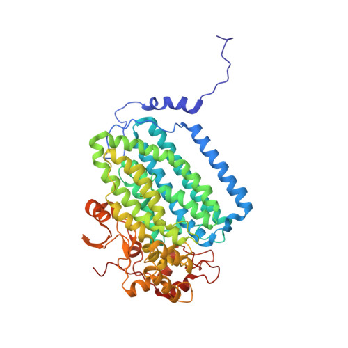
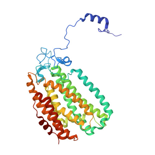
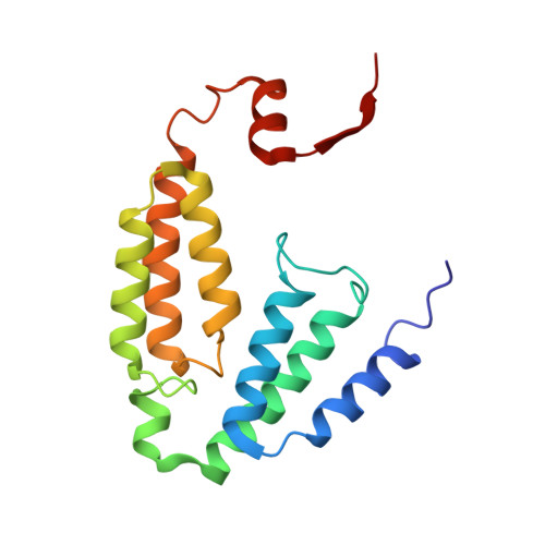
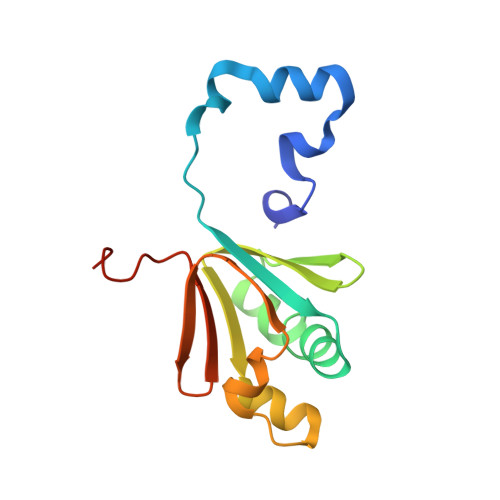
    Full activity of soluble methane monooxygenase (sMMO) depends upon the formation of a 1:1 complex of the regulatory protein MMOB with each alpha subunit of the (αβγ) 2 hydroxylase, sMMOH. Previous studies have shown that mutations in the core region of MMOB and in the N- and C-termini cause dramatic changes in the rate constants for steps in the sMMOH reaction cycle. Here, X-ray crystal structures are reported for the sMMOH complex with two double variants within the core region of MMOB, DBL1 (N107G/S110A), and DBL2 (S109A/T111A), as well as two variants in the MMOB N-terminal region, H33A and H5A. DBL1 causes a 150-fold decrease in the formation rate constant of the reaction cycle intermediate P , whereas DBL2 accelerates the reaction of the dinuclear Fe(IV) intermediate Q with substrates larger than methane by three- to fourfold. H33A also greatly slows P formation, while H5A modestly slows both formation of Q and its reactions with substrates. Complexation with DBL1 or H33A alters the position of sMMOH residue R245, which is part of a conserved hydrogen-bonding network encompassing the active site diiron cluster where P is formed. Accordingly, electron paramagnetic resonance spectra of sMMOH:DBL1 and sMMOH:H33A complexes differ markedly from that of sMMOH:MMOB, showing an altered electronic environment. In the sMMOH:DBL2 complex, the position of M247 in sMMOH is altered such that it enlarges a molecular tunnel associated with substrate entry into the active site. The H5A variant causes only subtle structural changes despite its kinetic effects, emphasizing the precise alignment of sMMOH and MMOB required for efficient catalysis.


  • Organizational Affiliation

    Department of Biochemistry, Molecular Biology, and Biophysics, University of Minnesota, Minneapolis, Minnesota 55455, United States.


Macromolecules
Find similar proteins by:  (by identity cutoff)  |  3D Structure
Entity ID: 1
MoleculeChains Sequence LengthOrganismDetailsImage
Methane monooxygenase component A alpha chain
A, E
515Methylosinus trichosporium OB3bMutation(s): 0 
Gene Names: CQW49_12480
UniProt
Find proteins for P27353 (Methylosinus trichosporium)
Explore P27353 
Go to UniProtKB:  P27353
Entity Groups  
Sequence Clusters30% Identity50% Identity70% Identity90% Identity95% Identity100% Identity
UniProt GroupP27353
Sequence Annotations
Expand
  • Reference Sequence
Find similar proteins by:  (by identity cutoff)  |  3D Structure
Entity ID: 2
MoleculeChains Sequence LengthOrganismDetailsImage
Methane monooxygenase beta chain
B, F
392Methylosinus trichosporium OB3bMutation(s): 0 
Gene Names: CQW49_12475
UniProt
Find proteins for A0A2D2D5X7 (Methylosinus trichosporium OB3b)
Explore A0A2D2D5X7 
Go to UniProtKB:  A0A2D2D5X7
Entity Groups  
Sequence Clusters30% Identity50% Identity70% Identity90% Identity95% Identity100% Identity
UniProt GroupA0A2D2D5X7
Sequence Annotations
Expand
  • Reference Sequence
Find similar proteins by:  (by identity cutoff)  |  3D Structure
Entity ID: 3
MoleculeChains Sequence LengthOrganismDetailsImage
Methane monooxygenase gamma chain
C, G
168Methylosinus trichosporium OB3bMutation(s): 0 
Gene Names: CQW49_12465
UniProt
Find proteins for A0A2D2D0T0 (Methylosinus trichosporium OB3b)
Explore A0A2D2D0T0 
Go to UniProtKB:  A0A2D2D0T0
Entity Groups  
Sequence Clusters30% Identity50% Identity70% Identity90% Identity95% Identity100% Identity
UniProt GroupA0A2D2D0T0
Sequence Annotations
Expand
  • Reference Sequence
Find similar proteins by:  (by identity cutoff)  |  3D Structure
Entity ID: 4
MoleculeChains Sequence LengthOrganismDetailsImage
Methane monooxygenase regulatory protein B
D, H
136Methylosinus trichosporium OB3bMutation(s): 1 
Gene Names: CQW49_12470
UniProt
Find proteins for P27356 (Methylosinus trichosporium)
Explore P27356 
Go to UniProtKB:  P27356
Entity Groups  
Sequence Clusters30% Identity50% Identity70% Identity90% Identity95% Identity100% Identity
UniProt GroupP27356
Sequence Annotations
Expand
  • Reference Sequence
Small Molecules
Ligands 3 Unique
IDChains Name / Formula / InChI Key2D Diagram3D Interactions
BEZ
Query on BEZ

Download Ideal Coordinates CCD File 
O [auth A],
RA [auth E]
BENZOIC ACID
C7 H6 O2
WPYMKLBDIGXBTP-UHFFFAOYSA-N
EDO
Query on EDO

Download Ideal Coordinates CCD File 
AA [auth B]
AB [auth F]
BA [auth B]
BB [auth F]
CA [auth B]
AA [auth B],
AB [auth F],
BA [auth B],
BB [auth F],
CA [auth B],
CB [auth F],
DA [auth B],
DB [auth F],
EA [auth B],
EB [auth F],
FA [auth B],
FB [auth F],
GA [auth C],
GB [auth F],
HA [auth C],
HB [auth G],
IA [auth C],
IB [auth G],
JA [auth D],
JB [auth G],
K [auth A],
KA [auth D],
KB [auth G],
L [auth A],
LA [auth D],
LB [auth H],
M [auth A],
N [auth A],
OA [auth E],
P [auth A],
PA [auth E],
Q [auth A],
QA [auth E],
R [auth A],
S [auth A],
SA [auth E],
T [auth A],
TA [auth E],
U [auth A],
UA [auth E],
V [auth A],
VA [auth E],
W [auth A],
WA [auth E],
X [auth A],
XA [auth E],
Y [auth A],
YA [auth F],
Z [auth B],
ZA [auth F]
1,2-ETHANEDIOL
C2 H6 O2
LYCAIKOWRPUZTN-UHFFFAOYSA-N
FE
Query on FE

Download Ideal Coordinates CCD File 
I [auth A],
J [auth A],
MA [auth E],
NA [auth E]
FE (III) ION
Fe
VTLYFUHAOXGGBS-UHFFFAOYSA-N
Experimental Data & Validation

Experimental Data

  • Method: X-RAY DIFFRACTION
  • Resolution: 1.82 Å
  • R-Value Free: 0.172 
  • R-Value Work: 0.146 
  • R-Value Observed: 0.147 
  • Space Group: P 21 21 21
Unit Cell:
Length ( Å )Angle ( ˚ )
a = 102.52α = 90
b = 105.6β = 90
c = 299.98γ = 90
Software Package:
Software NamePurpose
PHENIXrefinement
Aimlessdata scaling
PDB_EXTRACTdata extraction
XDSdata reduction
PHENIXphasing

Structure Validation

View Full Validation Report


Entry History & Funding Information

Deposition Data


Funding OrganizationLocationGrant Number
National Institutes of Health/National Institute of General Medical Sciences (NIH/NIGMS)United StatesGM118030
National Institutes of Health/National Institute of General Medical Sciences (NIH/NIGMS)United StatesGM118047
National Institutes of Health/National Institute of General Medical Sciences (NIH/NIGMS)United StatesGM08347

Revision History  (Full details and data files)

  • Version 1.0: 2021-12-29
    Type: Initial release
  • Version 1.1: 2022-01-19
    Changes: Database references
  • Version 1.2: 2023-10-18
    Changes: Data collection, Refinement description