7S5Q | pdb_00007s5q

Crystal structure of SARS-CoV-2 B.1.351 variant receptor binding domain in complex with neutralizing antibodies CS44 and COVA1-16


Experimental Data Snapshot

  • Method: X-RAY DIFFRACTION
  • Resolution: 2.88 Å
  • R-Value Free: 
    0.257 (Depositor), 0.254 (DCC) 
  • R-Value Work: 
    0.216 (Depositor), 0.213 (DCC) 
  • R-Value Observed: 
    0.218 (Depositor) 

Starting Model: experimental
View more details

wwPDB Validation 3D Report Full Report

Validation slider image for 7S5Q

Ligand Structure Quality Assessment 


This is version 1.2 of the entry. See complete history

Literature

SARS-CoV-2 Beta variant infection elicits potent lineage-specific and cross-reactive antibodies.

Reincke, S.M.Yuan, M.Kornau, H.C.Corman, V.M.van Hoof, S.Sanchez-Sendin, E.Ramberger, M.Yu, W.Hua, Y.Tien, H.Schmidt, M.L.Schwarz, T.Jeworowski, L.M.Brandl, S.E.Rasmussen, H.F.Homeyer, M.A.Stoffler, L.Barner, M.Kunkel, D.Huo, S.Horler, J.von Wardenburg, N.Kroidl, I.Eser, T.M.Wieser, A.Geldmacher, C.Hoelscher, M.Ganzer, H.Weiss, G.Schmitz, D.Drosten, C.Pruss, H.Wilson, I.A.Kreye, J.

(2022) Science 375: 782-787

  • DOI: https://doi.org/10.1126/science.abm5835
  • Primary Citation Related Structures: 
    7S5P, 7S5Q, 7S5R

  • PubMed Abstract: 

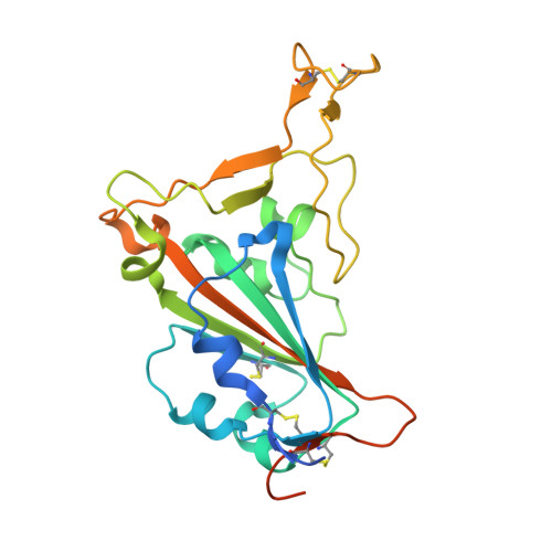
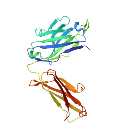
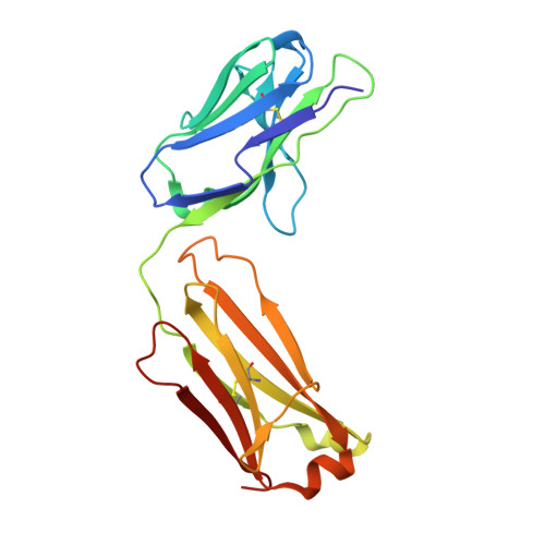
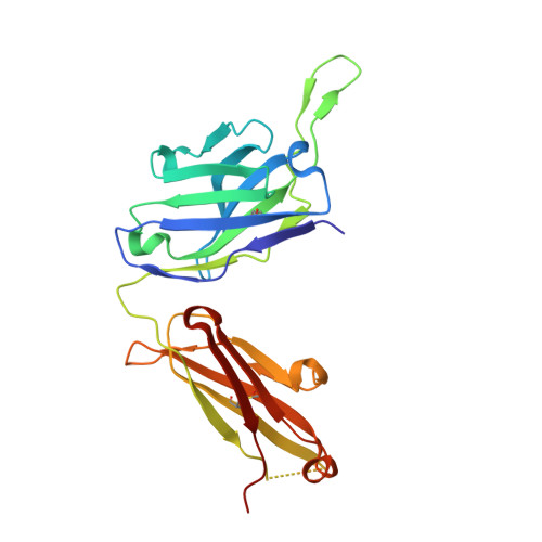
    Severe acute respiratory syndrome coronavirus 2 (SARS-CoV-2) Beta variant of concern (VOC) resists neutralization by major classes of antibodies from COVID-19 patients and vaccinated individuals. In this study, serum of Beta-infected patients revealed reduced cross-neutralization of wild-type virus. From these patients, we isolated Beta-specific and cross-reactive receptor-binding domain (RBD) antibodies. The Beta-specificity results from recruitment of VOC-specific clonotypes and accommodation of mutations present in Beta and Omicron into a major antibody class that is normally sensitive to these mutations. The Beta-elicited cross-reactive antibodies share genetic and structural features with wild type-elicited antibodies, including a public VH1-58 clonotype that targets the RBD ridge. These findings advance our understanding of the antibody response to SARS-CoV-2 shaped by antigenic drift, with implications for design of next-generation vaccines and therapeutics.


  • Organizational Affiliation
    • Department of Neurology and Experimental Neurology, Charité-Universitätsmedizin Berlin, corporate member of Freie Universität Berlin and Humboldt-Universität zu Berlin, Berlin, Germany.

Macromolecule Content 

  • Total Structure Weight: 123.69 kDa 
  • Atom Count: 8,323 
  • Modeled Residue Count: 1,068 
  • Deposited Residue Count: 1,118 
  • Unique protein chains: 5

Macromolecules

Find similar proteins by:|  3D Structure
Entity ID: 1
MoleculeChains  Sequence LengthOrganismDetailsImage
Spike protein S1231Severe acute respiratory syndrome coronavirus 2Mutation(s): 3 
Gene Names: S2
UniProt
Find proteins for P0DTC2 (Severe acute respiratory syndrome coronavirus 2)
Explore P0DTC2 
Go to UniProtKB:  P0DTC2
Entity Groups
Sequence Clusters30% Identity50% Identity70% Identity90% Identity95% Identity100% Identity
UniProt GroupP0DTC2
Glycosylation
Glycosylation Sites: 1Go to GlyGen: P0DTC2-1
Sequence Annotations
Expand
Reference Sequence
Find similar proteins by:|  3D Structure
Entity ID: 2
MoleculeChains  Sequence LengthOrganismDetailsImage
CS44 Heavy chain226Homo sapiensMutation(s): 0 
Find similar proteins by:|  3D Structure
Entity ID: 3
MoleculeChains  Sequence LengthOrganismDetailsImage
CS44 Light chain215Homo sapiensMutation(s): 0 
Find similar proteins by:|  3D Structure
Entity ID: 4
MoleculeChains  Sequence LengthOrganismDetailsImage
COVA1-16 Heavy chainD [auth H]232Homo sapiensMutation(s): 0 
Find similar proteins by:|  3D Structure
Entity ID: 5
MoleculeChains  Sequence LengthOrganismDetailsImage
COVA1-16 Light chainE [auth L]214Homo sapiensMutation(s): 0 

Small Molecules

Ligands 3 Unique
IDChains Name / Formula / InChI Key2D Diagram3D Interactions
NAG

Query on NAG


Download:Ideal Coordinates CCD File
F [auth A]2-acetamido-2-deoxy-beta-D-glucopyranose
C8 H15 N O6
OVRNDRQMDRJTHS-FMDGEEDCSA-N
SO4

Query on SO4


Download:Ideal Coordinates CCD File
I [auth A]
J [auth A]
K [auth A]
L [auth A]
M [auth A]
I [auth A],
J [auth A],
K [auth A],
L [auth A],
M [auth A],
N [auth A],
O [auth B],
Q [auth H],
R [auth H],
S [auth H],
V [auth L]
SULFATE ION
O4 S
QAOWNCQODCNURD-UHFFFAOYSA-L
EDO

Query on EDO


Download:Ideal Coordinates CCD File
G [auth A],
H [auth A],
P [auth H],
T [auth L],
U [auth L]
1,2-ETHANEDIOL
C2 H6 O2
LYCAIKOWRPUZTN-UHFFFAOYSA-N

Experimental Data & Validation

Experimental Data

  • Method: X-RAY DIFFRACTION
  • Resolution: 2.88 Å
  • R-Value Free:  0.257 (Depositor), 0.254 (DCC) 
  • R-Value Work:  0.216 (Depositor), 0.213 (DCC) 
  • R-Value Observed: 0.218 (Depositor) 
Space Group: P 21 21 21
Unit Cell:
Length ( Å )Angle ( ˚ )
a = 89.637α = 90
b = 120.97β = 90
c = 137.987γ = 90
Software Package:
Software NamePurpose
PHENIXrefinement
HKL-2000data scaling
PDB_EXTRACTdata extraction
HKL-2000data reduction
PHASERphasing

Structure Validation

View Full Validation Report


Ligand Structure Quality Assessment 


Entry History 

& Funding Information

Deposition Data


Funding OrganizationLocationGrant Number
Bill & Melinda Gates FoundationINV-004923
Bavarian State Ministry for Education, Culture, Science and Arts01KI20271

Revision History  (Full details and data files)

  • Version 1.0: 2022-03-09
    Type: Initial release
  • Version 1.1: 2023-10-18
    Changes: Data collection, Refinement description
  • Version 1.2: 2024-11-06
    Changes: Structure summary