Minimal Increments of Hydrophobic Collapse within the N-Terminus of the Neuropeptide Galanin.
Kraichely, K.N., Clinkscales, S.E., Hendy, C.M., Mendoza, E.A., Parnham, S., Giuliano, M.W.(2022) Biochemistry 61: 1151-1166
- PubMed: 35622960 Search on PubMed
- DOI: https://doi.org/10.1021/acs.biochem.2c00141
- Primary Citation Related Structures:
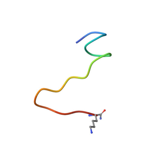
7S3O, 7S3Q, 7S3R - PubMed Abstract:
The neuropeptide galanin has a 35-year history as an intriguing target in drug design owing to its implication as a potential anticonvulsant and neuronal trophic factor among many other therapeutically interesting functions including analgesia and mood alteration. In this study, we report the structural characterization of three synthetic fragments of the galanin N-terminus in buffered aqueous solution: hGal(2-12)KK, hGal(1-12)KK, and hGal(1-17)KK. High-field two-dimensional 1 H- 1 H nuclear magnetic resonance (NMR) data were acquired for these fragments and used to derive distance restraints. We further utilized modified hydrogen bonding and dihedral restraints to reflect chemical shift patterns in the data, which revealed the signature of a weakly folded helix. Together, these sets of restraints were used to generate NMR structures of all three fragments, which depict a core of hydrophobic residues that cluster together regardless of the presence of a helical structure, and correspond to residues in the N-terminus of galanin that have been previously shown to be critical for binding its receptors. The helical structure only appears following the inclusion of Gly(1) in the sequence, and at longer sequence lengths, unlike many other peptides, the helix does not propagate. Rather, a few turns of poorly ordered helix appear to be a secondary consequence of clusters of hydrophobic sidechains that are conserved across all of the peptides in this study; the helices themselves appear ordered as a consequence of this clustering, and these clusters compare directly to those observed recently to make contacts between galanin and two of its receptor subtypes. Collapsed hydrophobic residues therefore organize and compose the functional core of human galanin and raise interesting questions about the nature of the conformational order in ligands that bind cell surface receptors.
- Department of Chemistry and Biochemistry, College of Charleston, Charleston, South Carolina 29424, United States.
Organizational Affiliation: