7S13 | pdb_00007s13

Crystal structure of Fab in complex with mouse CD96 dimer


Experimental Data Snapshot

  • Method: X-RAY DIFFRACTION
  • Resolution: 2.12 Å
  • R-Value Free: 
    0.265 (Depositor), 0.264 (DCC) 
  • R-Value Work: 
    0.217 (Depositor), 0.217 (DCC) 
  • R-Value Observed: 
    0.219 (Depositor) 

Starting Models: experimental
View more details

wwPDB Validation 3D Report Full Report

Validation slider image for 7S13

Ligand Structure Quality Assessment 


This is version 1.2 of the entry. See complete history

Literature

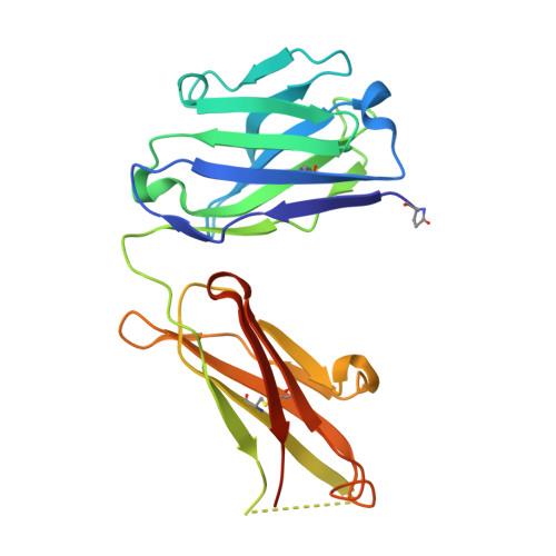
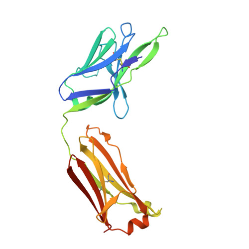
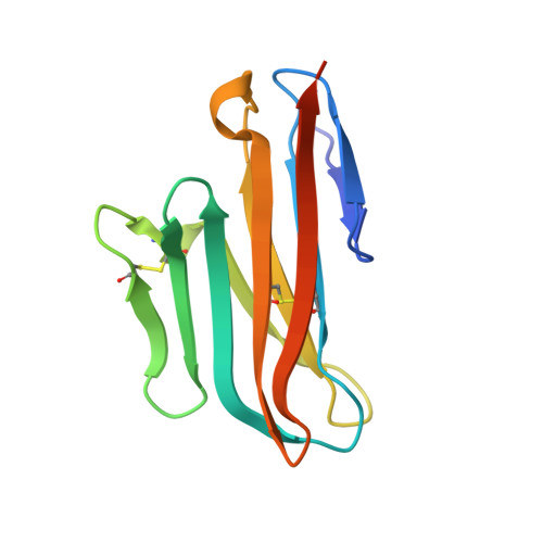
Antibody blockade of CD96 by distinct molecular mechanisms.

Lee, P.S.Chau, B.Barman, I.Bee, C.Jashnani, A.Hogan, J.M.Aguilar, B.Dollinger, G.Rajpal, A.Strop, P.

(2021) MAbs 13: 1979800-1979800

  • DOI: https://doi.org/10.1080/19420862.2021.1979800
  • Primary Citation Related Structures: 
    7S11, 7S13

  • PubMed Abstract: 

    The molecular interactions of mouse CD96 to CD155 ligand and to two surrogate antibodies have been investigated. Biophysical and structural studies demonstrate that CD96 forms a homodimer but assembles as 1:1 heterodimeric complexes with CD155 or with one of the surrogate antibodies, which compete for the same binding interface. In comparison, the other surrogate antibody binds across the mouse CD96 dimer and recognizes a quaternary epitope spanning both protomers to block exposure of the ligand-binding site. This study reveals different blocking mechanisms and modalities of these two antibodies and may provide insight into the functional effects of antibodies against CD96.


  • Organizational Affiliation
    • Discovery Biotherapeutics, Bristol Myers Squibb, Redwood City, CA, USA.

Macromolecule Content 

  • Total Structure Weight: 124.58 kDa 
  • Atom Count: 8,274 
  • Modeled Residue Count: 1,065 
  • Deposited Residue Count: 1,128 
  • Unique protein chains: 3

Macromolecules

Find similar proteins by:|  3D Structure
Entity ID: 1
MoleculeChains  Sequence LengthOrganismDetailsImage
Fab heavy chainA [auth H],
C [auth I]
226RattusMutation(s): 0 
Find similar proteins by:|  3D Structure
Entity ID: 2
MoleculeChains  Sequence LengthOrganismDetailsImage
Fab light chainB [auth L],
D [auth M]
213RattusMutation(s): 0 
Find similar proteins by:|  3D Structure
Entity ID: 3
MoleculeChains  Sequence LengthOrganismDetailsImage
T-cell surface protein tactileE [auth C],
F [auth D]
125Mus musculusMutation(s): 0 
Gene Names: Cd96
UniProt & NIH Common Fund Data Resources
Find proteins for Q3U0X8 (Mus musculus)
Explore Q3U0X8 
Go to UniProtKB:  Q3U0X8
IMPC:  MGI:1934368
Entity Groups
Sequence Clusters30% Identity50% Identity70% Identity90% Identity95% Identity100% Identity
UniProt GroupQ3U0X8
Glycosylation
Glycosylation Sites: 1Go to GlyGen: Q3U0X8-1
Sequence Annotations
Expand
Reference Sequence

Small Molecules

Modified Residues  1 Unique
IDChains TypeFormula2D DiagramParent
PCA
Query on PCA
A [auth H],
C [auth I]
L-PEPTIDE LINKINGC5 H7 N O3GLN

Experimental Data & Validation

Experimental Data

  • Method: X-RAY DIFFRACTION
  • Resolution: 2.12 Å
  • R-Value Free:  0.265 (Depositor), 0.264 (DCC) 
  • R-Value Work:  0.217 (Depositor), 0.217 (DCC) 
  • R-Value Observed: 0.219 (Depositor) 
Space Group: P 21 21 21
Unit Cell:
Length ( Å )Angle ( ˚ )
a = 52.03α = 90
b = 131.25β = 90
c = 186.87γ = 90
Software Package:
Software NamePurpose
PHENIXrefinement
XDSdata reduction
XSCALEdata scaling
PHASERphasing

Structure Validation

View Full Validation Report


Ligand Structure Quality Assessment 


Entry History 

& Funding Information

Deposition Data


Funding OrganizationLocationGrant Number
Not funded--

Revision History  (Full details and data files)

  • Version 1.0: 2021-10-20
    Type: Initial release
  • Version 1.1: 2023-10-18
    Changes: Data collection, Refinement description
  • Version 1.2: 2024-10-23
    Changes: Structure summary