7RW6

BORF2-APOBEC3Bctd Complex


Experimental Data Snapshot

  • Method: ELECTRON MICROSCOPY
  • Resolution: 2.55 Å
  • Aggregation State: PARTICLE 
  • Reconstruction Method: SINGLE PARTICLE 

wwPDB Validation   3D Report Full Report


This is version 1.1 of the entry. See complete history


Literature

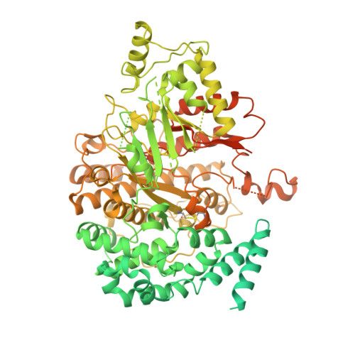
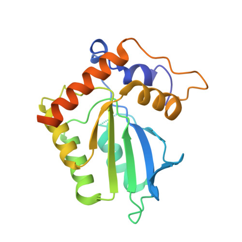
Cryo-EM structure of the EBV ribonucleotide reductase BORF2 and mechanism of APOBEC3B inhibition.

Shaban, N.M.Yan, R.Shi, K.Moraes, S.N.Cheng, A.Z.Carpenter, M.A.McLellan, J.S.Yu, Z.Harris, R.S.

(2022) Sci Adv 8: eabm2827-eabm2827

  • DOI: https://doi.org/10.1126/sciadv.abm2827
  • Primary Citation of Related Structures:  
    7RW6

  • PubMed Abstract: 

    Viruses use a plethora of mechanisms to evade immune responses. A recent example is neutralization of the nuclear DNA cytosine deaminase APOBEC3B by the Epstein-Barr virus (EBV) ribonucleotide reductase subunit BORF2. Cryo-EM studies of APOBEC3B-BORF2 complexes reveal a large >1000-Å 2 binding surface composed of multiple structural elements from each protein, which effectively blocks the APOBEC3B active site from accessing single-stranded DNA substrates. Evolutionary optimization is suggested by unique insertions in BORF2 absent from other ribonucleotide reductases and preferential binding to APOBEC3B relative to the highly related APOBEC3A and APOBEC3G enzymes. A molecular understanding of this pathogen-host interaction has potential to inform the development of drugs that block the interaction and liberate the natural antiviral activity of APOBEC3B. In addition, given a role for APOBEC3B in cancer mutagenesis, it may also be possible for information from the interaction to be used to develop DNA deaminase inhibitors.


  • Organizational Affiliation

    Department of Biochemistry, Molecular Biology, and Biophysics, Institute for Molecular Virology, Masonic Cancer Center, University of Minnesota, Minneapolis, MN 55455, USA.


Macromolecules
Find similar proteins by:  (by identity cutoff)  |  3D Structure
Entity ID: 1
MoleculeChains Sequence LengthOrganismDetailsImage
Maltose/maltodextrin-binding periplasmic protein,Ribonucleoside-diphosphate reductase large subunit
A, C
1,197Escherichia coli K-12human gammaherpesvirus 4
This entity is chimeric
Mutation(s): 5 
Gene Names: malEb4034JW3994RIR1BORF2
EC: 1.17.4.1
UniProt
Find proteins for P0AEX9 (Escherichia coli (strain K12))
Explore P0AEX9 
Go to UniProtKB:  P0AEX9
Find proteins for P03190 (Epstein-Barr virus (strain B95-8))
Explore P03190 
Go to UniProtKB:  P03190
Entity Groups  
Sequence Clusters30% Identity50% Identity70% Identity90% Identity95% Identity100% Identity
UniProt GroupsP03190P0AEX9
Sequence Annotations
Expand
  • Reference Sequence
Find similar proteins by:  (by identity cutoff)  |  3D Structure
Entity ID: 2
MoleculeChains Sequence LengthOrganismDetailsImage
DNA dC->dU-editing enzyme APOBEC-3B
B, D
215Homo sapiensMutation(s): 0 
Gene Names: APOBEC3B
EC: 3.5.4.38
UniProt & NIH Common Fund Data Resources
Find proteins for Q9UH17 (Homo sapiens)
Explore Q9UH17 
Go to UniProtKB:  Q9UH17
PHAROS:  Q9UH17
GTEx:  ENSG00000179750 
Entity Groups  
Sequence Clusters30% Identity50% Identity70% Identity90% Identity95% Identity100% Identity
UniProt GroupQ9UH17
Sequence Annotations
Expand
  • Reference Sequence
Experimental Data & Validation

Experimental Data

  • Method: ELECTRON MICROSCOPY
  • Resolution: 2.55 Å
  • Aggregation State: PARTICLE 
  • Reconstruction Method: SINGLE PARTICLE 

Structure Validation

View Full Validation Report


Entry History & Funding Information

Deposition Data


Funding OrganizationLocationGrant Number
National Institutes of Health/National Cancer Institute (NIH/NCI)United States--

Revision History  (Full details and data files)

  • Version 1.0: 2022-04-27
    Type: Initial release
  • Version 1.1: 2022-05-04
    Changes: Database references