7RTP

Structure of full-length human lambda-6A light chain JTO in complex with urea stabilizer 20 [1-(2-(7-(diethylamino)-4-methyl-2-oxo-2H-chromen-3-yl)ethyl)-3-(pyridin-3-ylmethyl)urea]


Experimental Data Snapshot

  • Method: X-RAY DIFFRACTION
  • Resolution: 2.09 Å
  • R-Value Free: 0.238 
  • R-Value Work: 0.179 
  • R-Value Observed: 0.182 

wwPDB Validation   3D Report Full Report


This is version 1.1 of the entry. See complete history


Literature

Amyloidogenic immunoglobulin light chain kinetic stabilizers comprising a simple urea linker module reveal a novel binding sub-site.

Yan, N.L.Nair, R.Chu, A.Wilson, I.A.Johnson, K.A.Morgan, G.J.Kelly, J.W.

(2022) Bioorg Med Chem Lett 60: 128571-128571

  • DOI: https://doi.org/10.1016/j.bmcl.2022.128571
  • Primary Citation of Related Structures:  
    7RTP

  • PubMed Abstract: 

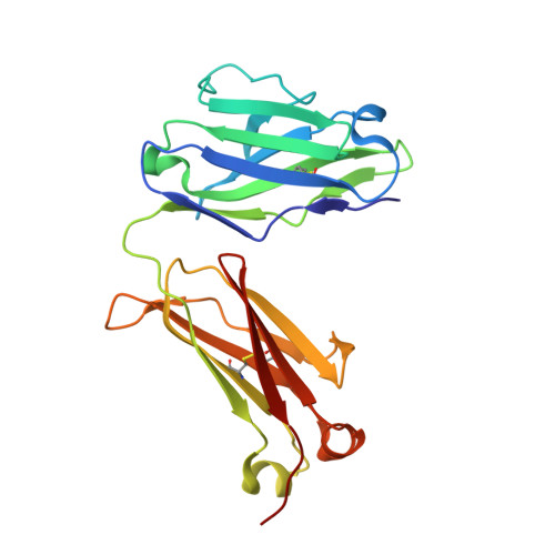
    In immunoglobulin light chain (LC) amyloidosis, the misfolding, or misfolding and misassembly of LC a protein or fragments thereof resulting from aberrant endoproteolysis, causes organ damage to patients. A small molecule "kinetic stabilizer" drug could slow or stop these processes and improve prognosis. We previously identified coumarin-based kinetic stabilizers of LCs that can be divided into four components, including a "linker module" and "distal substructure". Our prior studies focused on characterizing carbamate, hydantoin, and spirocyclic urea linker modules, which bind in a solvent-exposed site at the V L -V L domain interface of the LC dimer. Here, we report structure-activity relationship data on 7-diethylamino coumarin-based kinetic stabilizers. This substructure occupies the previously characterized "anchor cavity" and the "aromatic slit". The potencies of amide and urea linker modules terminating in a variety of distal substructures attached at the 3-position of this coumarin ring were assessed. Surprisingly, crystallographic data on a 7-diethylamino coumarin-based kinetic stabilizer reveals that the urea linker module and distal substructure attached at the 3-position bind a solvent-exposed region of the full-length LC dimer distinct from previously characterized sites. Our results further elaborate the small-molecule binding surface of LCs that could be occupied by potent and selective LC kinetic stabilizers.


  • Organizational Affiliation

    Department of Chemistry, The Scripps Research Institute, La Jolla, CA 92037, USA.


Macromolecules
Find similar proteins by:  (by identity cutoff)  |  3D Structure
Entity ID: 1
MoleculeChains Sequence LengthOrganismDetailsImage
JTO light chain
A, B
215Homo sapiensMutation(s): 0 
Entity Groups  
Sequence Clusters30% Identity50% Identity70% Identity90% Identity95% Identity100% Identity
Sequence Annotations
Expand
  • Reference Sequence
Small Molecules
Ligands 2 Unique
IDChains Name / Formula / InChI Key2D Diagram3D Interactions
NY9 (Subject of Investigation/LOI)
Query on NY9

Download Ideal Coordinates CCD File 
D [auth B]N-{2-[7-(diethylamino)-4-methyl-2-oxo-2H-1-benzopyran-3-yl]ethyl}-N'-[(pyridin-3-yl)methyl]urea
C23 H28 N4 O3
WLXNTQQMVXKPGD-UHFFFAOYSA-N
PO4
Query on PO4

Download Ideal Coordinates CCD File 
C [auth A],
E [auth B]
PHOSPHATE ION
O4 P
NBIIXXVUZAFLBC-UHFFFAOYSA-K
Experimental Data & Validation

Experimental Data

  • Method: X-RAY DIFFRACTION
  • Resolution: 2.09 Å
  • R-Value Free: 0.238 
  • R-Value Work: 0.179 
  • R-Value Observed: 0.182 
  • Space Group: P 21 21 21
Unit Cell:
Length ( Å )Angle ( ˚ )
a = 63.643α = 90
b = 80.585β = 90
c = 94.645γ = 90
Software Package:
Software NamePurpose
REFMACrefinement
PDB_EXTRACTdata extraction
XDSdata reduction
SCALAdata scaling
PHASERphasing

Structure Validation

View Full Validation Report


Entry History & Funding Information

Deposition Data


Funding OrganizationLocationGrant Number
National Institutes of Health/National Institute of Diabetes and Digestive and Kidney Disease (NIH/NIDDK)United StatesDK46335

Revision History  (Full details and data files)

  • Version 1.0: 2022-02-16
    Type: Initial release
  • Version 1.1: 2023-10-18
    Changes: Data collection, Refinement description