


The Functional Analysis of a Major Tyrosine Phosphorylation Site on Actin
Amelie, A., Dai, S., Shen, X., Horton, J.R., Zhang, X., Cheng, X.To be published.
Experimental Data Snapshot
Starting Model: experimental
View more details
wwPDB Validation 3D Report Full Report

Entity ID: 1 | |||||
|---|---|---|---|---|---|
| Molecule | Chains | Sequence Length | Organism | Details | Image |
| Phosphatidylinositol 3-kinase regulatory subunit beta | 114 | Homo sapiens | Mutation(s): 0 Gene Names: PIK3R2 | 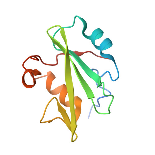 | |
UniProt & NIH Common Fund Data Resources | |||||
PHAROS: O00459 GTEx: ENSG00000105647 | |||||
Entity Groups | |||||
| Sequence Clusters | 30% Identity50% Identity70% Identity90% Identity95% Identity100% Identity | ||||
| UniProt Group | O00459 | ||||
Sequence AnnotationsExpand | |||||
Reference Sequence | |||||
Entity ID: 2 | |||||
|---|---|---|---|---|---|
| Molecule | Chains | Sequence Length | Organism | Details | Image |
| Actin, alpha skeletal muscle | 9 | Homo sapiens | Mutation(s): 0 EC: 3.6.4 | 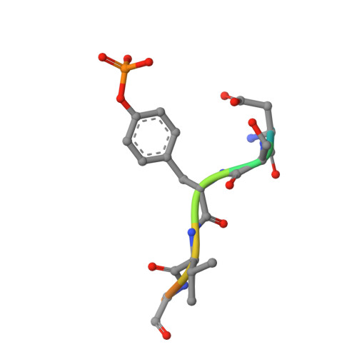 | |
UniProt & NIH Common Fund Data Resources | |||||
PHAROS: P68133 GTEx: ENSG00000143632 | |||||
Entity Groups | |||||
| UniProt Group | P68133 | ||||
Sequence AnnotationsExpand | |||||
Reference Sequence | |||||
| Ligands 1 Unique | |||||
|---|---|---|---|---|---|
| ID | Chains | Name / Formula / InChI Key | 2D Diagram | 3D Interactions | |
| EDO Download:Ideal Coordinates CCD File | I [auth A] J [auth A] K [auth A] L [auth C] M [auth C] | 1,2-ETHANEDIOL C2 H6 O2 LYCAIKOWRPUZTN-UHFFFAOYSA-N |  | ||
| Modified Residues 1 Unique | |||||
|---|---|---|---|---|---|
| ID | Chains | Type | Formula | 2D Diagram | Parent |
| PTR Query on PTR | B, D, F, H | L-PEPTIDE LINKING | C9 H12 N O6 P |  | TYR |
| Length ( Å ) | Angle ( ˚ ) |
|---|---|
| a = 188.083 | α = 90 |
| b = 41.432 | β = 100.294 |
| c = 65.875 | γ = 90 |
| Software Name | Purpose |
|---|---|
| PHENIX | refinement |
| HKL-2000 | data reduction |
| HKL-2000 | data scaling |
| PHENIX | phasing |
| Funding Organization | Location | Grant Number |
|---|---|---|
| National Institutes of Health/National Institute of General Medical Sciences (NIH/NIGMS) | United States | GM049245-23 |


