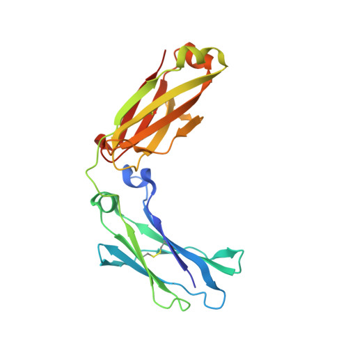
Effects of glycans and hinge on dynamics in the IgG1 Fc.
Bergonzo, C., Hoopes, J.T., Kelman, Z., Gallagher, D.T.(2023) J Biomol Struct Dyn : 1-9
- PubMed: 37897185
- DOI: https://doi.org/10.1080/07391102.2023.2270749
- Primary Citation of Related Structures:
7RHO - PubMed Abstract:
The crystallizable fragment (Fc) domain of immunoglobulin subclass IgG1 antibodies is engineered for a wide variety of pharmaceutical applications. Two important structural variables in Fc constructs are the hinge region connecting the Fc to the antigen binding fragments (Fab) and the glycans present in various glycoforms. These components affect receptor binding interactions that mediate immune activation. To design new antibody drugs, a robust in silico method for linking stability to structural changes is necessary. In this work, all-atom simulations were used to compare the dynamic behavior of the four structural variants arising from presence or absence of the hinge and glycans. We expressed the simplest of these constructs, the 'minimal Fc' with no hinge and no glycans, in Escherichia coli and report its crystal structure. The 'maximal Fc' that includes full hinge and G0F/G1F glycans is based on a previously reported structure, Protein Data Bank (PDB) ID: 5VGP. These, along with two intermediate structures (with only the glycans or with only the hinge) were used to independently measure the stability effects of the two structural variables using umbrella sampling simulations. Principal component analysis (PCA) was used to determine free energy effects along the Fc's dominant mode of motion. This work provides a comprehensive picture of the effects of hinge and glycans on Fc dynamics and stability.Communicated by Ramaswamy H. Sarma.
- Institute for Bioscience and Biotechnology Research, National Institute of Standards and Technology, The University of Maryland, Gudelsky Way, Rockville, MD, USA.
Organizational Affiliation:


