7RH2

IRF4 Transcription factor mutant -K59R


Experimental Data Snapshot

  • Method: X-RAY DIFFRACTION
  • Resolution: 2.47 Å
  • R-Value Free: 0.240 
  • R-Value Work: 0.203 
  • R-Value Observed: 0.205 

wwPDB Validation   3D Report Full Report


This is version 1.1 of the entry. See complete history


Literature

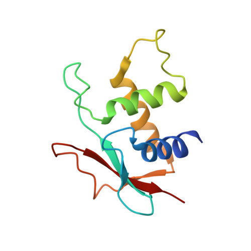
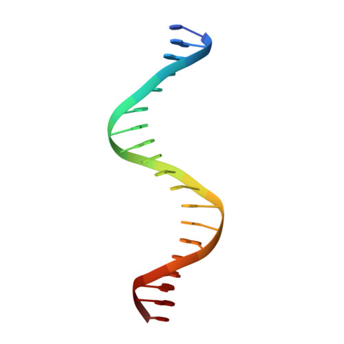
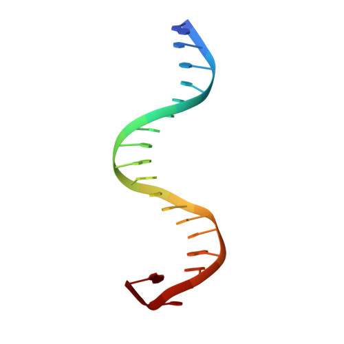
The molecular basis for the development of adult T-cell leukemia/lymphoma in patients with an IRF4 K59R mutation.

Sundararaj, S.Seneviratne, S.Williams, S.J.Enders, A.Casarotto, M.G.

(2022) Protein Sci 31: 787-796

  • DOI: https://doi.org/10.1002/pro.4260
  • Primary Citation of Related Structures:  
    7RH2

  • PubMed Abstract: 

    Interferon regulatory factor 4 (IRF4) is an essential regulator in the development of many immune cells, including B- and T-cells and has been implicated directly in numerous hematological malignancies, including adult T-cell leukemia/lymphoma (ATLL). Recently, an activating mutation in the DNA-binding domain of IRF4 (IRF4 K59R ) was found as a recurrent somatic mutation in ATLL patients. However, it remains unknown how this mutation gives rise to the observed oncogenic effect. To understand the mode of IRF4 K59R -mediated gain of function in ATLL pathogenesis, we have determined the structural and affinity basis of IRF4 K59R /DNA homodimer complex using X-ray crystallography and surface plasmon resonance. Our study shows that arginine substitution (R59) results in the reorientation of the side chain, enabling the guanidium group to interact with the phosphate backbone of the DNA helix. This markedly contrasts with the IRF4 WT wherein the K59 interacts exclusively with DNA bases. Further, the arginine mutation causes enhanced DNA bending, enabling the IRF4 K59R to interact more robustly with known DNA targets, as evidenced by increased binding affinity of the protein-DNA complex. Together, we demonstrate how key structural features underpin the basis for this activating mutation, thereby providing a molecular rationale for IRF4 K59R -mediated ATLL development.


  • Organizational Affiliation

    John Curtin School of Medical Research, Australian National University, Canberra, Australia.


Macromolecules

Find similar proteins by:  (by identity cutoff)  |  3D Structure
Entity ID: 1
MoleculeChains Sequence LengthOrganismDetailsImage
ICSAT transcription factor
A, B, G, H
111Homo sapiensMutation(s): 1 
UniProt & NIH Common Fund Data Resources
Find proteins for Q15306 (Homo sapiens)
Explore Q15306 
Go to UniProtKB:  Q15306
PHAROS:  Q15306
GTEx:  ENSG00000137265 
Entity Groups  
Sequence Clusters30% Identity50% Identity70% Identity90% Identity95% Identity100% Identity
UniProt GroupQ15306
Sequence Annotations
Expand
  • Reference Sequence

Find similar nucleic acids by:  Sequence   |   3D Structure  

Entity ID: 2
MoleculeChains LengthOrganismImage
DNA (5'-D(*CP*AP*AP*CP*TP*GP*AP*AP*AP*CP*CP*GP*AP*GP*AP*AP*AP*GP*C)-3')
C, D
19Homo sapiens
Sequence Annotations
Expand
  • Reference Sequence

Find similar nucleic acids by:  Sequence   |   3D Structure  

Entity ID: 3
MoleculeChains LengthOrganismImage
DNA (5'-D(*GP*CP*TP*TP*TP*CP*TP*CP*GP*GP*TP*TP*TP*CP*AP*GP*TP*TP*G)-3')
E, F
19Homo sapiens
Sequence Annotations
Expand
  • Reference Sequence
Experimental Data & Validation

Experimental Data

  • Method: X-RAY DIFFRACTION
  • Resolution: 2.47 Å
  • R-Value Free: 0.240 
  • R-Value Work: 0.203 
  • R-Value Observed: 0.205 
  • Space Group: P 31 2 1
Unit Cell:
Length ( Å )Angle ( ˚ )
a = 114.874α = 90
b = 114.874β = 90
c = 154.788γ = 120
Software Package:
Software NamePurpose
PHENIXrefinement
PHENIXrefinement
XDSdata reduction
Aimlessdata scaling
PHASERphasing

Structure Validation

View Full Validation Report


Entry History 

Deposition Data

Revision History  (Full details and data files)

  • Version 1.0: 2022-05-25
    Type: Initial release
  • Version 1.1: 2023-10-18
    Changes: Data collection, Refinement description