Investigation of the Molecular Details of the Interactions of Selenoglycosides and Human Galectin-3.
Raics, M., Balogh, A.K., Kishor, C., Timari, I., Medrano, F.J., Romero, A., Go, R.M., Blanchard, H., Szilagyi, L., E Kover, K., Feher, K.(2022) Int J Mol Sci 23
- PubMed: 35269646 Search on PubMedSearch on PubMed Central
- DOI: https://doi.org/10.3390/ijms23052494
- Primary Citation Related Structures:
7RDO, 7RDP - PubMed Abstract:
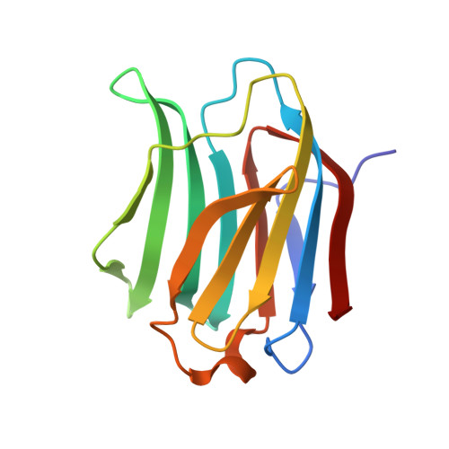
Human galectin-3 ( h Gal-3) is involved in a variety of biological processes and is implicated in wide range of diseases. As a result, targeting h Gal-3 for clinical applications has become an intense area of research. As a step towards the development of novel h Gal-3 inhibitors, we describe a study of the binding of two Se-containing h Gal-3 inhibitors, specifically that of di(β-D-galactopyranosyl)selenide (SeDG), in which two galactose rings are linked by one Se atom and a di(β-D-galactopyranosyl)diselenide (DSeDG) analogue with a diseleno bond between the two sugar units. The binding affinities of these derivatives to h Gal-3 were determined by 15 N- 1 H HSQC NMR spectroscopy and fluorescence anisotropy titrations in solution, indicating a slight decrease in the strength of interaction for SeDG compared to thiodigalactoside (TDG), a well-known inhibitor of h Gal-3, while DSeDG displayed a much weaker interaction strength. NMR and FA measurements showed that both seleno derivatives bind to the canonical S face site of h Gal-3 and stack against the conserved W181 residue also confirmed by X-ray crystallography, revealing canonical properties of the interaction. The interaction with DSeDG revealed two distinct binding modes in the crystal structure which are in fast exchange on the NMR time scale in solution, explaining a weaker interaction with h Gal-3 than SeDG. Using molecular dynamics simulations, we have found that energetic contributions to the binding enthalpies mainly differ in the electrostatic interactions and in polar solvation terms and are responsible for weaker binding of DSeDG compared to SeDG. Selenium-containing carbohydrate inhibitors of h Gal-3 showing canonical binding modes offer the potential of becoming novel hydrolytically stable scaffolds for a new class of h Gal-3 inhibitors.
- Molecular Recognition and Interaction Research Group, Hungarian Academy of Sciences, University of Debrecen, Egyetem tér 1, H-4032 Debrecen, Hungary.
Organizational Affiliation: