Discovery of an ʟ-amino acid ligase implicated in Staphylococcal sulfur amino acid metabolism.
Pederick, J.L., Horsfall, A.J., Jovcevski, B., Klose, J., Abell, A.D., Pukala, T.L., Bruning, J.B.(2022) J Biological Chem 298: 102392-102392
- PubMed: 35988643 Search on PubMedSearch on PubMed Central
- DOI: https://doi.org/10.1016/j.jbc.2022.102392
- Primary Citation Related Structures:
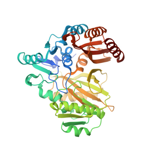
7R8P, 7R8Q - PubMed Abstract:
Enzymes involved in Staphylococcus aureus amino acid metabolism have recently gained traction as promising targets for the development of new antibiotics, however, not all aspects of this process are understood. The ATP-grasp superfamily includes enzymes that predominantly catalyze the ATP-dependent ligation of various carboxylate and amine substrates. One subset, ʟ-amino acid ligases (LALs), primarily catalyze the formation of dipeptide products in Gram-positive bacteria, however, their involvement in S. aureus amino acid metabolism has not been investigated. Here, we present the characterization of the putative ATP-grasp enzyme (SAOUHSC_02373) from S. aureus NCTC 8325 and its identification as a novel LAL. First, we interrogated the activity of SAOUHSC_02373 against a panel of ʟ-amino acid substrates. As a result, we identified SAOUHSC_02373 as an LAL with high selectivity for ʟ-aspartate and ʟ-methionine substrates, specifically forming an ʟ-aspartyl-ʟ-methionine dipeptide. Thus, we propose that SAOUHSC_02373 be assigned as ʟ-aspartate-ʟ-methionine ligase (LdmS). To further understand this unique activity, we investigated the mechanism of LdmS by X-ray crystallography, molecular modeling, and site-directed mutagenesis. Our results suggest that LdmS shares a similar mechanism to other ATP-grasp enzymes but possesses a distinctive active site architecture that confers selectivity for the ʟ-Asp and ʟ-Met substrates. Phylogenetic analysis revealed LdmS homologs are highly conserved in Staphylococcus and closely related Gram-positive Firmicutes. Subsequent genetic analysis upstream of the ldmS operon revealed several trans-acting regulatory elements associated with control of Met and Cys metabolism. Together, these findings support a role for LdmS in Staphylococcal sulfur amino acid metabolism.
- Institute for Photonics and Advanced Sensing (IPAS), School of Biological Sciences, The University of Adelaide, Adelaide, South Australia, Australia.
Organizational Affiliation: