7R6R

Crystal Structure of a Mycobacteriophage Cluster A2 Immunity Repressor:DNA Complex


Experimental Data Snapshot

  • Method: X-RAY DIFFRACTION
  • Resolution: 3.13 Å
  • R-Value Free: 0.248 
  • R-Value Work: 0.209 
  • R-Value Observed: 0.212 

wwPDB Validation   3D Report Full Report


This is version 1.2 of the entry. See complete history


Literature

A monomeric mycobacteriophage immunity repressor utilizes two domains to recognize an asymmetric DNA sequence.

McGinnis, R.J.Brambley, C.A.Stamey, B.Green, W.C.Gragg, K.N.Cafferty, E.R.Terwilliger, T.C.Hammel, M.Hollis, T.J.Miller, J.M.Gainey, M.D.Wallen, J.R.

(2022) Nat Commun 13: 4105-4105

  • DOI: https://doi.org/10.1038/s41467-022-31678-6
  • Primary Citation of Related Structures:  
    7R6R, 7TZ1

  • PubMed Abstract: 

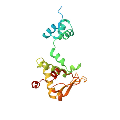
    Regulation of bacteriophage gene expression involves repressor proteins that bind and downregulate early lytic promoters. A large group of mycobacteriophages code for repressors that are unusual in also terminating transcription elongation at numerous binding sites (stoperators) distributed across the phage genome. Here we provide the X-ray crystal structure of a mycobacteriophage immunity repressor bound to DNA, which reveals the binding of a monomer to an asymmetric DNA sequence using two independent DNA binding domains. The structure is supported by small-angle X-ray scattering, DNA binding, molecular dynamics, and in vivo immunity assays. We propose a model for how dual DNA binding domains facilitate regulation of both transcription initiation and elongation, while enabling evolution of other superinfection immune specificities.


  • Organizational Affiliation

    Western Carolina University, Department of Chemistry and Physics, 111 Memorial Drive, Cullowhee, NC, 28723, USA.


Macromolecules

Find similar proteins by:  (by identity cutoff)  |  3D Structure
Entity ID: 1
MoleculeChains Sequence LengthOrganismDetailsImage
Immunity repressor203Mycobacterium phage TipsytheTRexMutation(s): 0 
Gene Names: SEA_TIPSYTHETREX_75
UniProt
Find proteins for A0A2D1GKF7 (Mycobacterium phage TipsytheTRex)
Explore A0A2D1GKF7 
Go to UniProtKB:  A0A2D1GKF7
Entity Groups  
Sequence Clusters30% Identity50% Identity70% Identity90% Identity95% Identity100% Identity
UniProt GroupA0A2D1GKF7
Sequence Annotations
Expand
  • Reference Sequence

Find similar nucleic acids by:  Sequence   |   3D Structure  

Entity ID: 2
MoleculeChains LengthOrganismImage
DNA (5'-D(P*TP*TP*TP*CP*GP*GP*TP*GP*GP*CP*TP*GP*TP*CP*AP*AP*GP*CP*GP*GP*G)-3')21Mycobacterium phage TipsytheTRex
Sequence Annotations
Expand
  • Reference Sequence

Find similar nucleic acids by:  Sequence   |   3D Structure  

Entity ID: 3
MoleculeChains LengthOrganismImage
DNA (5'-D(P*CP*CP*CP*GP*CP*TP*TP*GP*AP*CP*AP*GP*CP*CP*AP*CP*CP*GP*AP*AP*A)-3')21Mycobacterium phage TipsytheTRex
Sequence Annotations
Expand
  • Reference Sequence
Experimental Data & Validation

Experimental Data

  • Method: X-RAY DIFFRACTION
  • Resolution: 3.13 Å
  • R-Value Free: 0.248 
  • R-Value Work: 0.209 
  • R-Value Observed: 0.212 
  • Space Group: C 1 2 1
Unit Cell:
Length ( Å )Angle ( ˚ )
a = 132.49α = 90
b = 43.51β = 102.257
c = 89.28γ = 90
Software Package:
Software NamePurpose
PHENIXrefinement
REFMACrefinement
XDSdata reduction
XDSdata scaling
PHASERphasing
PHENIXmodel building

Structure Validation

View Full Validation Report


Entry History & Funding Information

Deposition Data


Funding OrganizationLocationGrant Number
Howard Hughes Medical Institute (HHMI)United States--

Revision History  (Full details and data files)

  • Version 1.0: 2022-07-20
    Type: Initial release
  • Version 1.1: 2022-07-27
    Changes: Database references
  • Version 1.2: 2023-10-18
    Changes: Data collection, Refinement description