Conserved Signal Transduction Mechanisms and Dark Recovery Kinetic Tuning in the Pseudomonadaceae Short Light, Oxygen, Voltage (LOV) Protein Family.
Arinkin, V., Granzin, J., Jaeger, K.E., Willbold, D., Krauss, U., Batra-Safferling, R.(2024) J Mol Biology : 168458-168458
- PubMed: 38280482 Search on PubMed
- DOI: https://doi.org/10.1016/j.jmb.2024.168458
- Primary Citation Related Structures:
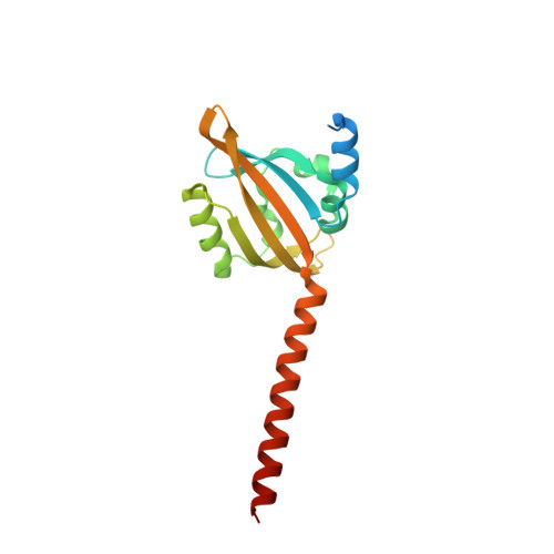
7R5N, 7YX0 - PubMed Abstract:
Light-Oxygen-Voltage (LOV) flavoproteins transduce a light signal into variable signaling outputs via a structural rearrangement in the sensory core domain, which is then relayed to fused effector domains via α-helical linker elements. Short LOV proteins from Pseudomonadaceae consist of a LOV sensory core and N- and C-terminal α-helices of variable length, providing a simple model system to study the molecular mechanism of allosteric activation. Here we report the crystal structures of two LOV proteins from Pseudomonas fluorescens - SBW25-LOV in the fully light-adapted state and Pf5-LOV in the dark-state. In a comparative analysis of the Pseudomonadaceae short LOVs, the structures demonstrate light-induced rotation of the core domains and splaying of the proximal A'α and Jα helices in the N and C-termini, highlighting evidence for a conserved signal transduction mechanism. Another distinguishing feature of the Pseudomonadaceae short LOV protein family is their highly variable dark recovery, ranging from seconds to days. Understanding this variability is crucial for tuning the signaling behavior of LOV-based optogenetic tools. At 37 °C, SBW25-LOV and Pf5-LOV exhibit adduct state lifetimes of 1470 min and 3.6 min, respectively. To investigate this remarkable difference in dark recovery rates, we targeted three residues lining the solvent channel entrance to the chromophore pocket where we introduced mutations by exchanging the non-conserved amino acids from SBW25-LOV into Pf5-LOV and vice versa. Dark recovery kinetics of the resulting mutants, as well as MD simulations and solvent cavity calculations on the crystal structures suggest a correlation between solvent accessibility and adduct lifetime.
- Institut für Biologische Informationsprozesse (IBI): Strukturbiochemie (IBI-7), Forschungszentrum Jülich, 52425 Jülich, Germany.
Organizational Affiliation: