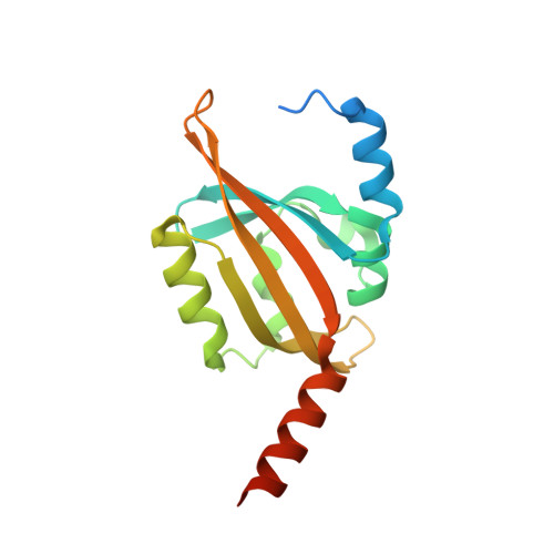
Residue alterations within a conserved hydrophobic pocket influence light, oxygen, voltage photoreceptor dark recovery.
Hemmer, S., Schulte, M., Knieps-Grunhagen, E., Granzin, J., Willbold, D., Jaeger, K.E., Batra-Safferling, R., Panwalkar, V., Krauss, U.(2023) Photochem Photobiol Sci 22: 713-727
- PubMed: 36480084 Search on PubMedSearch on PubMed Central
- DOI: https://doi.org/10.1007/s43630-022-00346-5
- Primary Citation Related Structures:
7R4S, 7R56 - PubMed Abstract:
Light, oxygen, voltage (LOV) photoreceptors are widely distributed throughout all kingdoms of life, and have in recent years, due to their modular nature, been broadly used as sensor domains for the construction of optogenetic tools. For understanding photoreceptor function as well as for optogenetic tool design and fine-tuning, a detailed knowledge of the photophysics, photochemistry, and structural changes underlying the LOV signaling paradigm is instrumental. Mutations that alter the lifetime of the photo-adduct signaling state represent a convenient handle to tune LOV sensor on/off kinetics and, thus, steady-state on/off equilibria of the photoreceptor (or optogenetic switch). Such mutations, however, should ideally only influence sensor kinetics, while being benign with regard to the nature of the structural changes that are induced by illumination, i.e., they should not result in a disruption of signal transduction. In the present study, we identify a conserved hydrophobic pocket for which mutations have a strong impact on the adduct-state lifetime across different LOV photoreceptor families. Using the slow cycling bacterial short LOV photoreceptor PpSB1-LOV, we show that the I48T mutation within this pocket, which accelerates adduct rupture, is otherwise structurally and mechanistically benign, i.e., light-induced structural changes, as probed by NMR spectroscopy and X-ray crystallography, are not altered in the variant. Additional mutations within the pocket of PpSB1-LOV and the introduction of homologous mutations in the LOV photoreceptor YtvA of Bacillus subtilis and the Avena sativa LOV2 domain result in similarly altered kinetics. Given the conserved nature of the corresponding structural region, the here identified mutations should find application in dark-recovery tuning of optogenetic tools and LOV photoreceptors, alike.
- Institut Für Molekulare Enzymtechnologie, Heinrich-Heine-Universität Düsseldorf, Forschungszentrum Jülich GmbH, 52425, Jülich, Germany.
Organizational Affiliation: