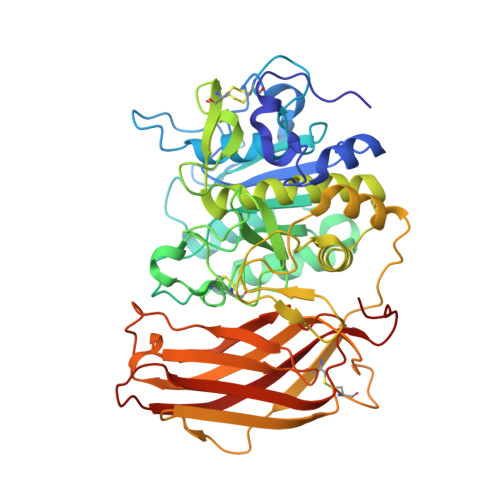
7QY0

X-ray structure of furin in complex with the dichlorophenylpyridine-based inhibitor 1


Experimental Data Snapshot

  • Method: X-RAY DIFFRACTION
  • Resolution: 1.54 Å
  • R-Value Free: 0.172 
  • R-Value Work: 0.149 
  • R-Value Observed: 0.150 

wwPDB Validation   3D Report Full Report


Ligand Structure Quality Assessment 


This is version 1.2 of the entry. See complete history


Literature

Dichlorophenylpyridine-Based Molecules Inhibit Furin through an Induced-Fit Mechanism.

Dahms, S.O.Schnapp, G.Winter, M.Buttner, F.H.Schleputz, M.Gnamm, C.Pautsch, A.Brandstetter, H.

(2022) ACS Chem Biol 17: 816-821

  • DOI: https://doi.org/10.1021/acschembio.2c00103
  • Primary Citation of Related Structures:  
    7QXY, 7QXZ, 7QY0, 7QY1, 7QY2

  • PubMed Abstract: 

    Inhibitors of the proprotein convertase furin might serve as broad-spectrum antiviral therapeutics. High cellular potency and antiviral activity against acute respiratory syndrome coronavirus 2 (SARS-CoV-2) have been reported for (3,5-dichlorophenyl)pyridine-derived furin inhibitors. Here we characterized the binding mechanism of this inhibitor class using structural, biophysical, and biochemical methods. We established a MALDI-TOF-MS-based furin activity assay, determined IC 50 values, and solved X-ray structures of (3,5-dichlorophenyl)pyridine-derived compounds in complex with furin. The inhibitors induced a substantial conformational rearrangement of the active-site cleft by exposing a central buried tryptophan residue. These changes formed an extended hydrophobic surface patch where the 3,5-dichlorophenyl moiety of the inhibitors was inserted into a newly formed binding pocket. Consistent with these structural rearrangements, we observed slow off-rate binding kinetics and strong structural stabilization in surface plasmon resonance and differential scanning fluorimetry experiments, respectively. The discovered furin conformation offers new opportunities for structure-based drug discovery.


  • Organizational Affiliation

    Department of Biosciences and Medical Biology, University of Salzburg, Hellbrunner Straße 34, A-5020 Salzburg, Austria.


Macromolecules
Find similar proteins by:  (by identity cutoff)  |  3D Structure
Entity ID: 1
MoleculeChains Sequence LengthOrganismDetailsImage
Furin480Homo sapiensMutation(s): 0 
Gene Names: FURINFURPACEPCSK3
EC: 3.4.21.75
UniProt & NIH Common Fund Data Resources
Find proteins for P09958 (Homo sapiens)
Explore P09958 
Go to UniProtKB:  P09958
PHAROS:  P09958
GTEx:  ENSG00000140564 
Entity Groups  
Sequence Clusters30% Identity50% Identity70% Identity90% Identity95% Identity100% Identity
UniProt GroupP09958
Sequence Annotations
Expand
  • Reference Sequence
Small Molecules
Ligands 6 Unique
IDChains Name / Formula / InChI Key2D Diagram3D Interactions
I0T (Subject of Investigation/LOI)
Query on I0T

Download Ideal Coordinates CCD File 
B [auth A]N-[[1-[[2-[3,5-bis(chloranyl)phenyl]-6-[6-[4-(2-methoxyethyl)piperazin-4-ium-1-yl]pyridin-3-yl]oxy-pyridin-4-yl]methyl]piperidin-1-ium-4-yl]methyl]ethanamide
C32 H42 Cl2 N6 O3
AUPHEGBHBGKBKL-UHFFFAOYSA-P
NAG
Query on NAG

Download Ideal Coordinates CCD File 
Q [auth A]2-acetamido-2-deoxy-beta-D-glucopyranose
C8 H15 N O6
OVRNDRQMDRJTHS-FMDGEEDCSA-N
DMS
Query on DMS

Download Ideal Coordinates CCD File 
K [auth A]
L [auth A]
M [auth A]
N [auth A]
O [auth A]
K [auth A],
L [auth A],
M [auth A],
N [auth A],
O [auth A],
P [auth A]
DIMETHYL SULFOXIDE
C2 H6 O S
IAZDPXIOMUYVGZ-UHFFFAOYSA-N
CA
Query on CA

Download Ideal Coordinates CCD File 
C [auth A],
D [auth A],
E [auth A]
CALCIUM ION
Ca
BHPQYMZQTOCNFJ-UHFFFAOYSA-N
CL
Query on CL

Download Ideal Coordinates CCD File 
J [auth A]CHLORIDE ION
Cl
VEXZGXHMUGYJMC-UHFFFAOYSA-M
NA
Query on NA

Download Ideal Coordinates CCD File 
F [auth A],
G [auth A],
H [auth A],
I [auth A]
SODIUM ION
Na
FKNQFGJONOIPTF-UHFFFAOYSA-N
Experimental Data & Validation

Experimental Data

  • Method: X-RAY DIFFRACTION
  • Resolution: 1.54 Å
  • R-Value Free: 0.172 
  • R-Value Work: 0.149 
  • R-Value Observed: 0.150 
  • Space Group: P 65 2 2
Unit Cell:
Length ( Å )Angle ( ˚ )
a = 131.447α = 90
b = 131.447β = 90
c = 155.329γ = 120
Software Package:
Software NamePurpose
PHENIXrefinement
PHENIXphasing
XDSdata reduction
XDSdata scaling

Structure Validation

View Full Validation Report


Ligand Structure Quality Assessment 


Entry History & Funding Information

Deposition Data


Funding OrganizationLocationGrant Number
Austrian Science FundAustriaM 2730

Revision History  (Full details and data files)

  • Version 1.0: 2022-04-20
    Type: Initial release
  • Version 1.1: 2022-04-27
    Changes: Database references
  • Version 1.2: 2024-01-31
    Changes: Data collection, Refinement description