7QUP

D. melanogaster 13-protofilament microtubule


Experimental Data Snapshot

  • Method: ELECTRON MICROSCOPY
  • Resolution: 3.80 Å
  • Aggregation State: FILAMENT 
  • Reconstruction Method: SINGLE PARTICLE 

wwPDB Validation   3D Report Full Report


This is version 1.2 of the entry. See complete history


Literature

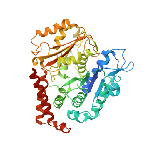
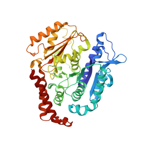
Diverse cytomotive actins and tubulins share a polymerization switch mechanism conferring robust dynamics.

Wagstaff, J.M.Planelles-Herrero, V.J.Sharov, G.Alnami, A.Kozielski, F.Derivery, E.Lowe, J.

(2023) Sci Adv 9: eadf3021-eadf3021

  • DOI: https://doi.org/10.1126/sciadv.adf3021
  • Primary Citation of Related Structures:  
    7QUC, 7QUD, 7QUP, 7QUQ

  • PubMed Abstract: 

    Protein filaments are used in myriads of ways to organize other molecules within cells. Some filament-forming proteins couple the hydrolysis of nucleotides to their polymerization cycle, thus powering the movement of other molecules. These filaments are termed cytomotive. Only members of the actin and tubulin protein superfamilies are known to form cytomotive filaments. We examined the basis of cytomotivity via structural studies of the polymerization cycles of actin and tubulin homologs from across the tree of life. We analyzed published data and performed structural experiments designed to disentangle functional components of these complex filament systems. Our analysis demonstrates the existence of shared subunit polymerization switches among both cytomotive actins and tubulins, i.e., the conformation of subunits switches upon assembly into filaments. These cytomotive switches can explain filament robustness, by enabling the coupling of kinetic and structural polarities required for cytomotive behaviors and by ensuring that single cytomotive filaments do not fall apart.


  • Organizational Affiliation

    MRC Laboratory of Molecular Biology, Francis Crick Avenue, Cambridge CB2 0QH, UK.


Macromolecules
Find similar proteins by:  (by identity cutoff)  |  3D Structure
Entity ID: 1
MoleculeChains Sequence LengthOrganismDetailsImage
Tubulin alpha-1 chain475Drosophila melanogasterMutation(s): 0 
Gene Names: alphaTub84BtubA84BCG1913
UniProt
Find proteins for P06603 (Drosophila melanogaster)
Explore P06603 
Go to UniProtKB:  P06603
Entity Groups  
Sequence Clusters30% Identity50% Identity70% Identity90% Identity95% Identity100% Identity
UniProt GroupP06603
Sequence Annotations
Expand
  • Reference Sequence
Find similar proteins by:  (by identity cutoff)  |  3D Structure
Entity ID: 2
MoleculeChains Sequence LengthOrganismDetailsImage
Tubulin beta-1 chain425Drosophila melanogasterMutation(s): 0 
UniProt
Find proteins for Q24560 (Drosophila melanogaster)
Explore Q24560 
Go to UniProtKB:  Q24560
Entity Groups  
Sequence Clusters30% Identity50% Identity70% Identity90% Identity95% Identity100% Identity
UniProt GroupQ24560
Sequence Annotations
Expand
  • Reference Sequence
Small Molecules
Ligands 3 Unique
IDChains Name / Formula / InChI Key2D Diagram3D Interactions
GTP
Query on GTP

Download Ideal Coordinates CCD File 
AF [auth 8C]
BC [auth 11E]
BD [auth 2A]
CE [auth 5C]
DC [auth 12A]
AF [auth 8C],
BC [auth 11E],
BD [auth 2A],
CE [auth 5C],
DC [auth 12A],
DF [auth 8E],
ED [auth 2C],
FE [auth 5E],
FF [auth 9A],
GC [auth 12C],
HD [auth 2E],
HE [auth 6A],
IF [auth 9C],
JC [auth 12E],
JD [auth 3A],
KE [auth 6C],
LC [auth 13A],
LF [auth 9E],
MD [auth 3C],
NB [auth 10A],
NE [auth 6E],
OC [auth 13C],
PD [auth 3E],
PE [auth 7A],
QB [auth 10C],
RC [auth 13E],
RD [auth 4A],
SE [auth 7C],
TB [auth 10E],
TC [auth 1A],
UD [auth 4C],
VB [auth 11A],
VE [auth 7E],
WC [auth 1C],
XD [auth 4E],
XE [auth 8A],
YB [auth 11C],
ZC [auth 1E],
ZD [auth 5A]
GUANOSINE-5'-TRIPHOSPHATE
C10 H16 N5 O14 P3
XKMLYUALXHKNFT-UUOKFMHZSA-N
GDP
Query on GDP

Download Ideal Coordinates CCD File 
AC [auth 11D]
BE [auth 5B]
CF [auth 8D]
DD [auth 2B]
EE [auth 5D]
AC [auth 11D],
BE [auth 5B],
CF [auth 8D],
DD [auth 2B],
EE [auth 5D],
FC [auth 12B],
GD [auth 2D],
HF [auth 9B],
IC [auth 12D],
JE [auth 6B],
KF [auth 9D],
LD [auth 3B],
ME [auth 6D],
NC [auth 13B],
OD [auth 3D],
PB [auth 10B],
QC [auth 13D],
RE [auth 7B],
SB [auth 10D],
TD [auth 4B],
UE [auth 7D],
VC [auth 1B],
WD [auth 4D],
XB [auth 11B],
YC [auth 1D],
ZE [auth 8B]
GUANOSINE-5'-DIPHOSPHATE
C10 H15 N5 O11 P2
QGWNDRXFNXRZMB-UUOKFMHZSA-N
MG
Query on MG

Download Ideal Coordinates CCD File 
AD [auth 1E]
AE [auth 5A]
BF [auth 8C]
CC [auth 11E]
CD [auth 2A]
AD [auth 1E],
AE [auth 5A],
BF [auth 8C],
CC [auth 11E],
CD [auth 2A],
DE [auth 5C],
EC [auth 12A],
EF [auth 8E],
FD [auth 2C],
GE [auth 5E],
GF [auth 9A],
HC [auth 12C],
ID [auth 2E],
IE [auth 6A],
JF [auth 9C],
KC [auth 12E],
KD [auth 3A],
LE [auth 6C],
MC [auth 13A],
MF [auth 9E],
ND [auth 3C],
OB [auth 10A],
OE [auth 6E],
PC [auth 13C],
QD [auth 3E],
QE [auth 7A],
RB [auth 10C],
SC [auth 13E],
SD [auth 4A],
TE [auth 7C],
UB [auth 10E],
UC [auth 1A],
VD [auth 4C],
WB [auth 11A],
WE [auth 7E],
XC [auth 1C],
YD [auth 4E],
YE [auth 8A],
ZB [auth 11C]
MAGNESIUM ION
Mg
JLVVSXFLKOJNIY-UHFFFAOYSA-N
Experimental Data & Validation

Experimental Data

  • Method: ELECTRON MICROSCOPY
  • Resolution: 3.80 Å
  • Aggregation State: FILAMENT 
  • Reconstruction Method: SINGLE PARTICLE 

Structure Validation

View Full Validation Report


Entry History & Funding Information

Deposition Data


Funding OrganizationLocationGrant Number
Wellcome TrustUnited Kingdom202754/Z/16/Z
Medical Research Council (MRC, United Kingdom)United KingdomU105184326

Revision History  (Full details and data files)

  • Version 1.0: 2022-11-02
    Type: Initial release
  • Version 1.1: 2023-04-19
    Changes: Database references, Refinement description
  • Version 1.2: 2023-10-04
    Changes: Data collection, Refinement description