7QST

Crystal structure of homing endonuclease-associated PhoVMA intein (C1A)


Experimental Data Snapshot

  • Method: X-RAY DIFFRACTION
  • Resolution: 2.49 Å
  • R-Value Free: 0.243 
  • R-Value Work: 0.183 
  • R-Value Observed: 0.186 

wwPDB Validation   3D Report Full Report


This is version 1.3 of the entry. See complete history


Literature

Structural Basis for the Propagation of Homing Endonuclease-Associated Inteins.

Beyer, H.M.Iwai, H.

(2022) Front Mol Biosci 9: 855511-855511

  • DOI: https://doi.org/10.3389/fmolb.2022.855511
  • Primary Citation of Related Structures:  
    7QSS, 7QST, 7QSU

  • PubMed Abstract: 

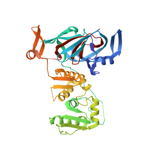
    Inteins catalyze their removal from a host protein through protein splicing. Inteins that contain an additional site-specific endonuclease domain display genetic mobility via a process termed "homing" and thereby act as selfish DNA elements. We elucidated the crystal structures of two archaeal inteins associated with an active or inactive homing endonuclease domain. This analysis illustrated structural diversity in the accessory domains (ACDs) associated with the homing endonuclease domain. To augment homing endonucleases with highly specific DNA cleaving activity using the intein scaffold, we engineered the ACDs and characterized their homing site recognition. Protein engineering of the ACDs in the inteins illuminated a possible strategy for how inteins could avoid their extinction but spread via the acquisition of a diverse accessory domain.


  • Organizational Affiliation

    Institute of Biotechnology, University of Helsinki, Helsinki, Finland.


Macromolecules
Find similar proteins by:  (by identity cutoff)  |  3D Structure
Entity ID: 1
MoleculeChains Sequence LengthOrganismDetailsImage
V-type ATP synthase subunit A379Pyrococcus horikoshiiMutation(s): 1 
Gene Names: HA331_07965
UniProt
Find proteins for O57728 (Pyrococcus horikoshii (strain ATCC 700860 / DSM 12428 / JCM 9974 / NBRC 100139 / OT-3))
Explore O57728 
Go to UniProtKB:  O57728
Entity Groups  
Sequence Clusters30% Identity50% Identity70% Identity90% Identity95% Identity100% Identity
UniProt GroupO57728
Sequence Annotations
Expand
  • Reference Sequence
Experimental Data & Validation

Experimental Data

  • Method: X-RAY DIFFRACTION
  • Resolution: 2.49 Å
  • R-Value Free: 0.243 
  • R-Value Work: 0.183 
  • R-Value Observed: 0.186 
  • Space Group: C 1 2 1
Unit Cell:
Length ( Å )Angle ( ˚ )
a = 91.08α = 90
b = 81.87β = 112.85
c = 67.69γ = 90
Software Package:
Software NamePurpose
PHENIXrefinement
XDSdata reduction
XDSdata scaling
Auto-Rickshawphasing

Structure Validation

View Full Validation Report


Entry History & Funding Information

Deposition Data


Funding OrganizationLocationGrant Number
Academy of FinlandDenmark137995, 277335
Novo Nordisk FoundationDenmarkNNF17OC0025402, NNF17OC0027550

Revision History  (Full details and data files)

  • Version 1.0: 2022-03-30
    Type: Initial release
  • Version 1.1: 2022-04-06
    Changes: Data collection, Structure summary
  • Version 1.2: 2022-04-13
    Changes: Database references
  • Version 1.3: 2024-01-31
    Changes: Data collection, Refinement description