7QP1 | pdb_00007qp1

Crystal structure of metacaspase from candida glabrata with calcium


Experimental Data Snapshot

  • Method: X-RAY DIFFRACTION
  • Resolution: 3.00 Å
  • R-Value Free: 
    0.261 (Depositor), 0.257 (DCC) 
  • R-Value Work: 
    0.208 (Depositor), 0.212 (DCC) 
  • R-Value Observed: 
    0.210 (Depositor) 

Starting Model: experimental
View more details

wwPDB Validation 3D Report Full Report

Validation slider image for 7QP1

This is version 1.1 of the entry. See complete history

Literature

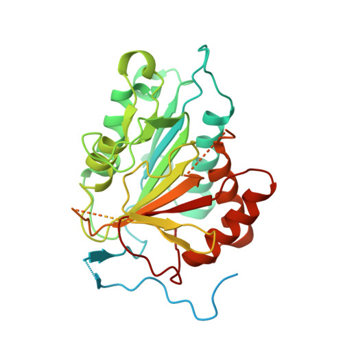
Structural and molecular determinants of Candida glabrata metacaspase maturation and activation by calcium.

Conchou, L.Doumeche, B.Galisson, F.Violot, S.Dugelay, C.Diesis, E.Page, A.Bienvenu, A.L.Picot, S.Aghajari, N.Ballut, L.

(2022) Commun Biol 5: 1158-1158

  • DOI: https://doi.org/10.1038/s42003-022-04091-4
  • Primary Citation Related Structures: 
    7QP0, 7QP1

  • PubMed Abstract: 

    Metacaspases are caspase-like homologs which undergo a complex maturation process involving multiple intra-chain cleavages resulting in a composite enzyme made of a p10 and a p20 domain. Their proteolytic activity involving a cysteine-histidine catalytic dyad, show peptide bond cleavage specificity in the C-terminal to lysine and arginine, with both maturation- and catalytic processes being calcium-dependent. Here, we present the structure of a metacaspase from the yeast Candida glabrata, CgMCA-I, in complex with a unique calcium along with a structure in which three magnesium ions are bound. We show that the Ca 2+ ion interacts with a loop in the vicinity of the catalytic site. The reorganization of this cation binding loop, by bringing together the two catalytic residues, could be one of the main structural determinants triggering metacaspase activation. Enzymatic exploration of CgMCA-I confirmed that the maturation process implies a trans mechanism with sequential cleavages.


  • Organizational Affiliation
    • Molecular Microbiology and Structural Biochemistry, UMR 5086, CNRS-Université de Lyon, F-69367, Lyon, France.

Macromolecule Content 

  • Total Structure Weight: 87.84 kDa 
  • Atom Count: 4,025 
  • Modeled Residue Count: 510 
  • Deposited Residue Count: 784 
  • Unique protein chains: 1

Macromolecules

Find similar proteins by:|  3D Structure
Entity ID: 1
MoleculeChains  Sequence LengthOrganismDetailsImage
Metacaspase-1
A, B
392Nakaseomyces glabratusMutation(s): 0 
Gene Names: MCA1CAGL0I10945g
EC: 3.4.22
UniProt
Find proteins for Q6FPX9 (Candida glabrata (strain ATCC 2001 / BCRC 20586 / JCM 3761 / NBRC 0622 / NRRL Y-65 / CBS 138))
Explore Q6FPX9 
Go to UniProtKB:  Q6FPX9
Entity Groups
Sequence Clusters30% Identity50% Identity70% Identity90% Identity95% Identity100% Identity
UniProt GroupQ6FPX9
Sequence Annotations
Expand
Reference Sequence

Experimental Data & Validation

Experimental Data

  • Method: X-RAY DIFFRACTION
  • Resolution: 3.00 Å
  • R-Value Free:  0.261 (Depositor), 0.257 (DCC) 
  • R-Value Work:  0.208 (Depositor), 0.212 (DCC) 
  • R-Value Observed: 0.210 (Depositor) 
Space Group: P 32
Unit Cell:
Length ( Å )Angle ( ˚ )
a = 97.37α = 90
b = 97.37β = 90
c = 54.6γ = 120
Software Package:
Software NamePurpose
PHENIXrefinement
PDB_EXTRACTdata extraction
XDSdata reduction
XSCALEdata scaling
PHASERphasing

Structure Validation

View Full Validation Report


Entry History 

& Funding Information

Deposition Data


Funding OrganizationLocationGrant Number
Centre National de la Recherche Scientifique (CNRS)France--

Revision History  (Full details and data files)

  • Version 1.0: 2023-01-11
    Type: Initial release
  • Version 1.1: 2024-01-31
    Changes: Data collection, Refinement description