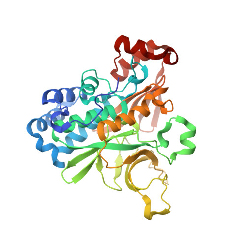
Fusarium verticillioides NAT1 (FDB2) N-malonyltransferase is structurally, functionally and phylogenetically distinct from its N-acetyltransferase (NAT) homologues.
Karagianni, E.P., Kontomina, E., Lowe, E.D., Athanasopoulos, K., Papanikolaou, G., Garefalaki, V., Kotseli, V., Zaliou, S., Grimaud, T., Arvaniti, K., Tsatiri, M.A., Fakis, G., Glenn, A.E., Roversi, P., Abuhammad, A., Ryan, A., Sim, R.B., Sim, E., Boukouvala, S.(2023) FEBS J 290: 2412-2436
- PubMed: 36178468 Search on PubMed
- DOI: https://doi.org/10.1111/febs.16642
- Primary Citation Related Structures:
7QI3 - PubMed Abstract:
Fusarium endophytes damage cereal crops and contaminate produce with mycotoxins. Those fungi overcome the main chemical defence of host via detoxification by a malonyl-CoA-dependent enzyme homologous to xenobiotic metabolizing arylamine N-acetyltransferase (NAT). In Fusarium verticillioides (teleomorph Gibberella moniliformis, GIBMO), this N-malonyltransferase activity is attributed to (GIBMO)NAT1, and the fungus has two additional isoenzymes, (GIBMO)NAT3 (N-acetyltransferase) and (GIBMO)NAT2 (unknown function). We present the crystallographic structure of (GIBMO)NAT1, also modelling other fungal NAT homologues. Monomeric (GIBMO)NAT1 is distinctive, with access to the catalytic core through two "tunnel-like" entries separated by a "bridge-like" helix. In the quaternary arrangement, (GIBMO)NAT1 monomers interact in pairs along an extensive interface whereby one entry of each monomer is covered by the N-terminus of the other monomer. Although monomeric (GIBMO)NAT1 apparently accommodates acetyl-CoA better than malonyl-CoA, dimerization changes the active site to allow malonyl-CoA to reach the catalytic triad (Cys110, His158 and Asp173) via the single uncovered entry, and anchor its terminal carboxyl-group via hydrogen bonds to Arg109, Asn157 and Thr261. Lacking a terminal carboxyl-group, acetyl-CoA cannot form such stabilizing interactions, while longer acyl-CoAs enter the active site but cannot reach catalytic Cys. Other NAT isoenzymes lack such structural features, with (GIBMO)NAT3 resembling bacterial NATs and (GIBMO)NAT2 adopting a structure intermediate between (GIBMO)NAT1 and (GIBMO)NAT3. Biochemical assays confirmed differential donor substrate preference of (GIBMO)NAT isoenzymes, with phylogenetic analysis demonstrating evolutionary separation. Given the role of (GIBMO)NAT1 in enhancing Fusarium pathogenicity, unravelling the structure and function of this enzyme may benefit research into more targeted strategies for pathogen control.
- Department of Molecular Biology and Genetics, Democritus University of Thrace, Alexandroupolis, Greece.
Organizational Affiliation: