7QGV | pdb_00007qgv

Solid-state NMR structure of Teixobactin-Lipid II.


Experimental Data Snapshot

  • Method: SOLID-STATE NMR
  • Conformers Calculated: 25 
  • Conformers Submitted: 25 
  • Selection Criteria: all calculated structures submitted 

wwPDB Validation 3D Report Full Report

Validation slider image for 7QGV

This is version 2.0 of the entry. See complete history

Literature

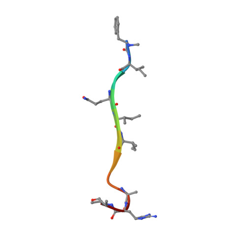
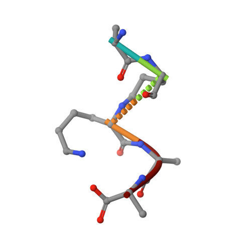
Teixobactin kills bacteria by a two-pronged attack on the cell envelope.

Shukla, R.Lavore, F.Maity, S.Derks, M.G.N.Jones, C.R.Vermeulen, B.J.A.Melcrova, A.Morris, M.A.Becker, L.M.Wang, X.Kumar, R.Medeiros-Silva, J.van Beekveld, R.A.M.Bonvin, A.M.J.J.Lorent, J.H.Lelli, M.Nowick, J.S.MacGillavry, H.D.Peoples, A.J.Spoering, A.L.Ling, L.L.Hughes, D.E.Roos, W.H.Breukink, E.Lewis, K.Weingarth, M.

(2022) Nature 608: 390-396

  • DOI: https://doi.org/10.1038/s41586-022-05019-y
  • Primary Citation Related Structures: 
    7QGV

  • PubMed Abstract: 

    Antibiotics that use novel mechanisms are needed to combat antimicrobial resistance 1-3 . Teixobactin 4 represents a new class of antibiotics with a unique chemical scaffold and lack of detectable resistance. Teixobactin targets lipid II, a precursor of peptidoglycan 5 . Here we unravel the mechanism of teixobactin at the atomic level using a combination of solid-state NMR, microscopy, in vivo assays and molecular dynamics simulations. The unique enduracididine C-terminal headgroup of teixobactin specifically binds to the pyrophosphate-sugar moiety of lipid II, whereas the N terminus coordinates the pyrophosphate of another lipid II molecule. This configuration favours the formation of a β-sheet of teixobactins bound to the target, creating a supramolecular fibrillar structure. Specific binding to the conserved pyrophosphate-sugar moiety accounts for the lack of resistance to teixobactin 4 . The supramolecular structure compromises membrane integrity. Atomic force microscopy and molecular dynamics simulations show that the supramolecular structure displaces phospholipids, thinning the membrane. The long hydrophobic tails of lipid II concentrated within the supramolecular structure apparently contribute to membrane disruption. Teixobactin hijacks lipid II to help destroy the membrane. Known membrane-acting antibiotics also damage human cells, producing undesirable side effects. Teixobactin damages only membranes that contain lipid II, which is absent in eukaryotes, elegantly resolving the toxicity problem. The two-pronged action against cell wall synthesis and cytoplasmic membrane produces a highly effective compound targeting the bacterial cell envelope. Structural knowledge of the mechanism of teixobactin will enable the rational design of improved drug candidates.


  • Organizational Affiliation
    • NMR Spectroscopy, Bijvoet Centre for Biomolecular Research, Department of Chemistry, Faculty of Science, Utrecht University, Utrecht, The Netherlands.

Macromolecule Content 

  • Total Structure Weight: 10.66 kDa 
  • Atom Count: 708 
  • Modeled Residue Count: 64 
  • Deposited Residue Count: 64 
  • Unique protein chains: 2

Macromolecules

Find similar proteins by:  Sequence   |   3D Structure  
Entity ID: 1
MoleculeChains  Sequence LengthOrganismDetailsImage
Teixobactin
A, B, C, D
11Eleftheria terraeMutation(s): 0 
Find similar proteins by:  Sequence   |   3D Structure  
Entity ID: 2
MoleculeChains  Sequence LengthOrganismDetailsImage
Lipid II
E, F, G, H
5Staphylococcus aureusMutation(s): 0 

Oligosaccharides

Help  
Entity ID: 3
MoleculeChains Length2D Diagram GlycosylationD Interactions
2-acetamido-2-deoxy-beta-D-glucopyranose-(1-4)-N-acetyl-alpha-muramic acid
I, J, K, L
2N/A
Glycosylation Resources
GlyTouCan: G88499BX
GlyCosmos: G88499BX

Small Molecules

Ligands 2 Unique
IDChains Name / Formula / InChI Key2D Diagram3D Interactions
P1W

Query on P1W


Download:Ideal Coordinates CCD File
BA [auth C]
EA [auth D]
FA [auth D]
O [auth A]
P [auth A]
BA [auth C],
EA [auth D],
FA [auth D],
O [auth A],
P [auth A],
Q [auth A],
R [auth B],
U [auth B],
V [auth B],
W [auth C],
X [auth C],
Y [auth C]
3-methylbut-2-en-1-ol
C5 H10 O
ASUAYTHWZCLXAN-UHFFFAOYSA-N
2PO

Query on 2PO


Download:Ideal Coordinates CCD File
AA [auth C]
CA [auth D]
DA [auth D]
M [auth A]
N [auth A]
AA [auth C],
CA [auth D],
DA [auth D],
M [auth A],
N [auth A],
S [auth B],
T [auth B],
Z [auth C]
PHOSPHONATE
H O3 P
ABLZXFCXXLZCGV-UHFFFAOYSA-L
Modified Residues  1 Unique
IDChains TypeFormula2D DiagramParent
28J
Query on 28J
A, B, C, D
D-PEPTIDE LINKINGC6 H13 N O2

--

DGN
Query on DGN
A, B, C, D
D-PEPTIDE LINKINGC5 H10 N2 O3

--

DTH
Query on DTH
A, B, C, D
D-PEPTIDE LINKINGC4 H9 N O3

--

EI4
Query on EI4
A, B, C, D
L-PEPTIDE LINKINGC6 H12 N4 O2ARG
ZAE
Query on ZAE
A, B, C, D
D-PEPTIDE LINKINGC10 H13 N O2

--

DAL
Query on DAL
E, F, G, H
D-PEPTIDE LINKINGC3 H7 N O2

--

DGL
Query on DGL
E, F, G, H
D-PEPTIDE LINKINGC5 H9 N O4

--

Biologically Interesting Molecules (External Reference) 

3 Unique

Experimental Data & Validation

Experimental Data

  • Method: SOLID-STATE NMR
  • Conformers Calculated: 25 
  • Conformers Submitted: 25 
  • Selection Criteria: all calculated structures submitted 

Structure Validation

View Full Validation Report


Entry History 

& Funding Information

Deposition Data


Funding OrganizationLocationGrant Number
Netherlands Organisation for Scientific Research (NWO)Netherlands--

Revision History  (Full details and data files)

  • Version 1.0: 2022-08-03
    Type: Initial release
  • Version 1.1: 2022-09-21
    Changes: Database references
  • Version 1.2: 2023-05-17
    Changes: Structure summary
  • Version 1.3: 2023-06-14
    Changes: Other
  • Version 2.0: 2023-11-15
    Changes: Advisory, Atomic model, Data collection, Derived calculations