Combined Structural and Computational Study of the mRubyFT Fluorescent Timer Locked in Its Blue Form.
Boyko, K.M., Khrenova, M.G., Nikolaeva, A.Y., Dorovatovskii, P.V., Vlaskina, A.V., Subach, O.M., Popov, V.O., Subach, F.V.(2023) Int J Mol Sci 24
- PubMed: 37175610 Search on PubMedSearch on PubMed Central
- DOI: https://doi.org/10.3390/ijms24097906
- Primary Citation Related Structures:
7Q6B - PubMed Abstract:
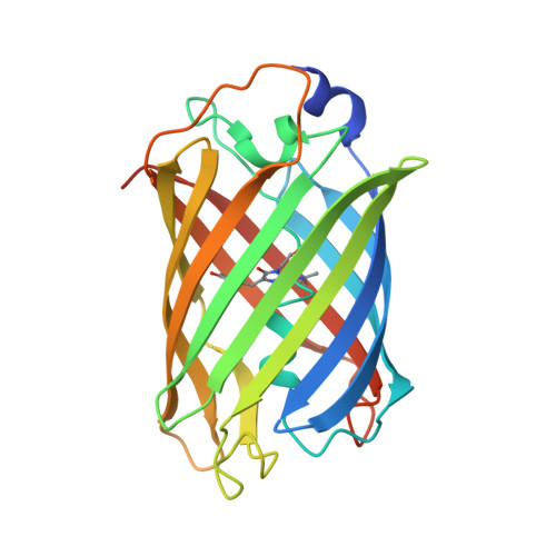
The mRubyFT is a monomeric genetically encoded fluorescent timer based on the mRuby2 fluorescent protein, which is characterized by the complete maturation of the blue form with the subsequent conversion to the red one. It has higher brightness in mammalian cells and higher photostability compared with other fluorescent timers. A high-resolution structure is a known characteristic of the mRubyFT with the red form chromophore, but structural details of its blue form remain obscure. In order to obtain insight into this, we obtained an S148I variant of the mRubyFT (mRubyFT S148I ) with the blocked over time blue form of the chromophore. X-ray data at a 1.8 Å resolution allowed us to propose a chromophore conformation and its interactions with the neighboring residues. The imidazolidinone moiety of the chromophore is completely matured, being a conjugated π-system. The methine bridge is not oxidized in the blue form bringing flexibility to the phenolic moiety that manifests itself in poor electron density. Integration of these data with the results of molecular dynamic simulation disclosed that the OH group of the phenolic moiety forms a hydrogen bond with the side chain of the T163 residue. A detailed comparison of mRubyFT S148I with other available structures of the blue form of fluorescent proteins, Blue102 and mTagBFP, revealed a number of characteristic differences. Molecular dynamic simulations with the combined quantum mechanic/molecular mechanic potentials demonstrated that the blue form exists in two protonation states, anion and zwitterion, both sharing enolate tautomeric forms of the C=C-O - fragment. These two forms have similar excitation energies, as evaluated by calculations. Finally, excited state molecular dynamic simulations showed that excitation of the chromophore in both protonation states leads to the same anionic fluorescent state. The data obtained shed light on the structural features and spectral properties of the blue form of the mRubyFT timer.
- Bach Institute of Biochemistry, Research Center of Biotechnology of the Russian Academy of Sciences, Leninsky Prospekt. 33, bld. 2, 119071 Moscow, Russia.
Organizational Affiliation: