Characterization of a phylogenetically distinct extradiol dioxygenase involved in the bacterial catabolism of lignin-derived aromatic compounds.
Navas, L.E., Zahn, M., Bajwa, H., Grigg, J.C., Wolf, M.E., Chan, A.C.K., Murphy, M.E.P., McGeehan, J.E., Eltis, L.D.(2022) J Biological Chem 298: 101871-101871
- PubMed: 35346686 Search on PubMedSearch on PubMed Central
- DOI: https://doi.org/10.1016/j.jbc.2022.101871
- Primary Citation Related Structures:
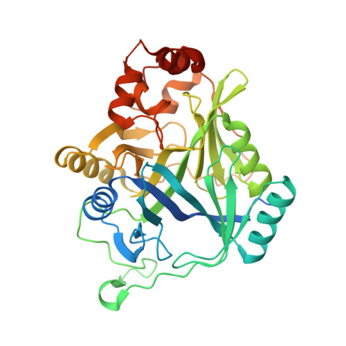
7Q2A - PubMed Abstract:
The actinobacterium Rhodococcus jostii RHA1 grows on a remarkable variety of aromatic compounds and has been studied for applications ranging from the degradation of polychlorinated biphenyls to the valorization of lignin, an underutilized component of biomass. In RHA1, the catabolism of two classes of lignin-derived compounds, alkylphenols and alkylguaiacols, involves a phylogenetically distinct extradiol dioxygenase, AphC, previously misannotated as BphC, an enzyme involved in biphenyl catabolism. To better understand the role of AphC in RHA1 catabolism, we first showed that purified AphC had highest apparent specificity for 4-propylcatechol (k cat /K M ∼10 6 M -1 s -1 ), and its apparent specificity for 4-alkylated substrates followed the trend for alkylguaiacols: propyl > ethyl > methyl > phenyl > unsubstituted. We also show AphC only poorly cleaved 3-phenylcatechol, the preferred substrate of BphC. Moreover, AphC and BphC cleaved 3-phenylcatechol and 4-phenylcatechol with different regiospecificities, likely due to the substrates' binding mode. A crystallographic structure of the AphC·4-ethylcatechol binary complex to 1.59 Å resolution revealed that the catechol is bound to the active site iron in a bidentate manner and that the substrate's alkyl side chain is accommodated by a hydrophobic pocket. Finally, we show RHA1 grows on a mixture of 4-ethylguaiacol and guaiacol, simultaneously catabolizing these substrates through meta-cleavage and ortho-cleavage pathways, respectively, suggesting that the specificity of AphC helps to prevent the routing of catechol through the Aph pathway. Overall, this study contributes to our understanding of the bacterial catabolism of aromatic compounds derived from lignin, and the determinants of specificity in extradiol dioxygenases.
- Department of Microbiology and Immunology, Life Sciences Institute, The University of British Columbia, Vancouver, Canada.
Organizational Affiliation: