7Q13

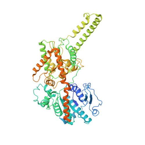
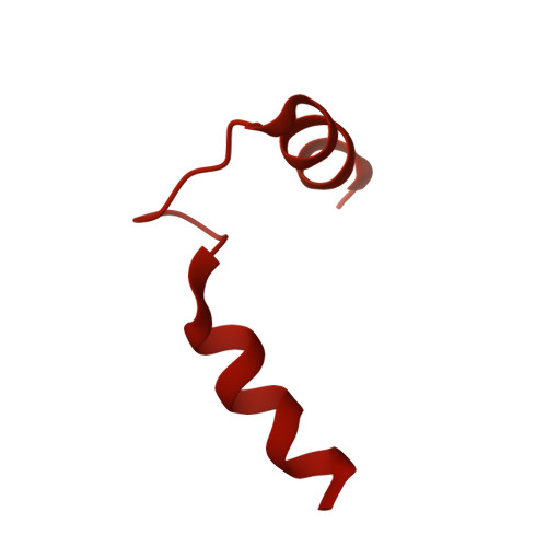
Human GYS1-GYG1 complex activated state bound to glucose-6-phosphate, uridine diphosphate, and glucose


Experimental Data Snapshot

  • Method: ELECTRON MICROSCOPY
  • Resolution: 3.00 Å
  • Aggregation State: PARTICLE 
  • Reconstruction Method: SINGLE PARTICLE 

Starting Models: experimental
View more details

wwPDB Validation   3D Report Full Report


This is version 1.1 of the entry. See complete history


Literature

Molecular basis for the regulation of human glycogen synthase by phosphorylation and glucose-6-phosphate.

McCorvie, T.J.Loria, P.M.Tu, M.Han, S.Shrestha, L.Froese, D.S.Ferreira, I.M.Berg, A.P.Yue, W.W.

(2022) Nat Struct Mol Biol 29: 628-638

  • DOI: https://doi.org/10.1038/s41594-022-00799-3
  • Primary Citation of Related Structures:  
    7Q0B, 7Q0S, 7Q12, 7Q13

  • PubMed Abstract: 

    Glycogen synthase (GYS1) is the central enzyme in muscle glycogen biosynthesis. GYS1 activity is inhibited by phosphorylation of its amino (N) and carboxyl (C) termini, which is relieved by allosteric activation of glucose-6-phosphate (Glc6P). We present cryo-EM structures at 3.0-4.0 Å resolution of phosphorylated human GYS1, in complex with a minimal interacting region of glycogenin, in the inhibited, activated and catalytically competent states. Phosphorylations of specific terminal residues are sensed by different arginine clusters, locking the GYS1 tetramer in an inhibited state via intersubunit interactions. The Glc6P activator promotes conformational change by disrupting these interactions and increases the flexibility of GYS1, such that it is poised to adopt a catalytically competent state when the sugar donor UDP-glucose (UDP-glc) binds. We also identify an inhibited-like conformation that has not transitioned into the activated state, in which the locking interaction of phosphorylation with the arginine cluster impedes subsequent conformational changes due to Glc6P binding. Our results address longstanding questions regarding the mechanism of human GYS1 regulation.


  • Organizational Affiliation

    Centre for Medicines Discovery, Nuffield Department of Clinical Medicine, University of Oxford, Oxford, UK.


Macromolecules
Find similar proteins by:  (by identity cutoff)  |  3D Structure
Entity ID: 1
MoleculeChains Sequence LengthOrganismDetailsImage
Glycogen [starch] synthase, muscleA,
F [auth B],
G [auth C],
H [auth D]
737Homo sapiensMutation(s): 0 
Gene Names: GYS1GYS
EC: 2.4.1.11
UniProt & NIH Common Fund Data Resources
Find proteins for P13807 (Homo sapiens)
Explore P13807 
Go to UniProtKB:  P13807
PHAROS:  P13807
GTEx:  ENSG00000104812 
Entity Groups  
Sequence Clusters30% Identity50% Identity70% Identity90% Identity95% Identity100% Identity
UniProt GroupP13807
Sequence Annotations
Expand
  • Reference Sequence
Find similar proteins by:  (by identity cutoff)  |  3D Structure
Entity ID: 2
MoleculeChains Sequence LengthOrganismDetailsImage
Glycogenin-1B [auth E],
C [auth F],
D [auth G],
E [auth H]
350Homo sapiensMutation(s): 0 
Gene Names: GYG1GYG
EC: 2.4.1.186
UniProt & NIH Common Fund Data Resources
Find proteins for P46976 (Homo sapiens)
Explore P46976 
Go to UniProtKB:  P46976
PHAROS:  P46976
GTEx:  ENSG00000163754 
Entity Groups  
Sequence Clusters30% Identity50% Identity70% Identity90% Identity95% Identity100% Identity
UniProt GroupP46976
Sequence Annotations
Expand
  • Reference Sequence
Small Molecules
Ligands 3 Unique
IDChains Name / Formula / InChI Key2D Diagram3D Interactions
UDP (Subject of Investigation/LOI)
Query on UDP

Download Ideal Coordinates CCD File 
J [auth A],
M [auth B],
P [auth C],
S [auth D]
URIDINE-5'-DIPHOSPHATE
C9 H14 N2 O12 P2
XCCTYIAWTASOJW-XVFCMESISA-N
G6P (Subject of Investigation/LOI)
Query on G6P

Download Ideal Coordinates CCD File 
I [auth A],
L [auth B],
O [auth C],
R [auth D]
6-O-phosphono-alpha-D-glucopyranose
C6 H13 O9 P
NBSCHQHZLSJFNQ-DVKNGEFBSA-N
GLC (Subject of Investigation/LOI)
Query on GLC

Download Ideal Coordinates CCD File 
K [auth A],
N [auth B],
Q [auth C],
T [auth D]
alpha-D-glucopyranose
C6 H12 O6
WQZGKKKJIJFFOK-DVKNGEFBSA-N
Experimental Data & Validation

Experimental Data

  • Method: ELECTRON MICROSCOPY
  • Resolution: 3.00 Å
  • Aggregation State: PARTICLE 
  • Reconstruction Method: SINGLE PARTICLE 
EM Software:
TaskSoftware PackageVersion
MODEL REFINEMENTPHENIX
RECONSTRUCTIONRELION3.1.1.

Structure Validation

View Full Validation Report


Entry History & Funding Information

Deposition Data


Funding OrganizationLocationGrant Number
Other private--

Revision History  (Full details and data files)

  • Version 1.0: 2022-07-27
    Type: Initial release
  • Version 1.1: 2024-07-17
    Changes: Data collection, Refinement description