7PZZ

Crystal structure of serine hydroxymethyltransferase, isoform 2 from Arabidopsis thaliana (SHM2)


Experimental Data Snapshot

  • Method: X-RAY DIFFRACTION
  • Resolution: 1.65 Å
  • R-Value Free: 0.178 
  • R-Value Work: 0.158 
  • R-Value Observed: 0.158 

wwPDB Validation   3D Report Full Report


This is version 1.1 of the entry. See complete history


Literature

Arabidopsis thaliana serine hydroxymethyltransferases: functions, structures, and perspectives.

Nogues, I.Sekula, B.Angelaccio, S.Grzechowiak, M.Tramonti, A.Contestabile, R.Ruszkowski, M.

(2022) Plant Physiol Biochem 187: 37-49

  • DOI: https://doi.org/10.1016/j.plaphy.2022.07.025
  • Primary Citation of Related Structures:  
    7PZZ, 7Q00, 7QPE, 7QX8

  • PubMed Abstract: 

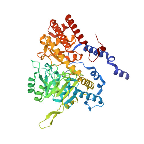
    Serine hydroxymethyltransferase (SHM) is one of the hallmarks of one-carbon metabolism. In plants, isoforms of SHM participate in photorespiration and/or transfer the one-carbon unit from L-serine to tetrahydrofolate (THF), hence producing 5,10-CH 2 -THF that is needed, e.g., for biosynthesis of methionine, thymidylate, and purines. These links highlight the importance of SHM activity in DNA biogenesis, its epigenetic methylations, and in stress responses. Plant genomes encode several SHM isoforms that localize to cytosol, mitochondria, plastids, and nucleus. In this work, we present a thorough functional and structural characterization of all seven SHM isoforms from Arabidopsis thaliana (AtSHM1-7). In particular, we analyzed tissue-specific expression profiles of the AtSHM genes. We also compared catalytic properties of the active AtSHM1-4 in terms of catalytic efficiency in both directions and inhibition by the THF substrate. Despite numerous attempts to rescue the SHM activity of AtSHM5-7, we failed, which points towards different physiological functions of these isoforms. Comparative analysis of experimental and predicted three-dimensional structures of AtSHM1-7 proteins indicated differences in regions that surround the entrance to the active site cavity.


  • Organizational Affiliation

    Research Institute on Terrestrial Ecosystems, Italian National Research Council, Monterotondo Scalo, Rome, Italy.


Macromolecules
Find similar proteins by:  (by identity cutoff)  |  3D Structure
Entity ID: 1
MoleculeChains Sequence LengthOrganismDetailsImage
Serine hydroxymethyltransferase 2, mitochondrial
A, B, C, D
480Arabidopsis thalianaMutation(s): 0 
Gene Names: SHM2SHMT2At5g26780F2P16.40
EC: 2.1.2.1
UniProt
Find proteins for Q94C74 (Arabidopsis thaliana)
Explore Q94C74 
Go to UniProtKB:  Q94C74
Entity Groups  
Sequence Clusters30% Identity50% Identity70% Identity90% Identity95% Identity100% Identity
UniProt GroupQ94C74
Sequence Annotations
Expand
  • Reference Sequence
Small Molecules
Ligands 5 Unique
IDChains Name / Formula / InChI Key2D Diagram3D Interactions
EPE
Query on EPE

Download Ideal Coordinates CCD File 
M [auth B]4-(2-HYDROXYETHYL)-1-PIPERAZINE ETHANESULFONIC ACID
C8 H18 N2 O4 S
JKMHFZQWWAIEOD-UHFFFAOYSA-N
PEG
Query on PEG

Download Ideal Coordinates CCD File 
LA [auth D],
X [auth C]
DI(HYDROXYETHYL)ETHER
C4 H10 O3
MTHSVFCYNBDYFN-UHFFFAOYSA-N
SO4
Query on SO4

Download Ideal Coordinates CCD File 
DA [auth C],
EA [auth C],
FA [auth C]
SULFATE ION
O4 S
QAOWNCQODCNURD-UHFFFAOYSA-L
EDO
Query on EDO

Download Ideal Coordinates CCD File 
AA [auth C]
BA [auth C]
CA [auth C]
E [auth A]
F [auth A]
AA [auth C],
BA [auth C],
CA [auth C],
E [auth A],
F [auth A],
G [auth A],
H [auth A],
HA [auth D],
I [auth A],
IA [auth D],
J [auth A],
JA [auth D],
KA [auth D],
L [auth B],
MA [auth D],
N [auth B],
NA [auth D],
O [auth B],
OA [auth D],
P [auth B],
Q [auth B],
R [auth B],
T [auth B],
U [auth C],
V [auth C],
W [auth C],
Y [auth C],
Z [auth C]
1,2-ETHANEDIOL
C2 H6 O2
LYCAIKOWRPUZTN-UHFFFAOYSA-N
CL
Query on CL

Download Ideal Coordinates CCD File 
GA [auth C],
K [auth A],
PA [auth D],
S [auth B]
CHLORIDE ION
Cl
VEXZGXHMUGYJMC-UHFFFAOYSA-M
Modified Residues  1 Unique
IDChains TypeFormula2D DiagramParent
LLP
Query on LLP
A, B, C, D
L-PEPTIDE LINKINGC14 H22 N3 O7 PLYS
Experimental Data & Validation

Experimental Data

  • Method: X-RAY DIFFRACTION
  • Resolution: 1.65 Å
  • R-Value Free: 0.178 
  • R-Value Work: 0.158 
  • R-Value Observed: 0.158 
  • Space Group: P 21 21 21
Unit Cell:
Length ( Å )Angle ( ˚ )
a = 115.201α = 90
b = 131.236β = 90
c = 151.389γ = 90
Software Package:
Software NamePurpose
PHENIXrefinement
XDSdata reduction
PHASERphasing

Structure Validation

View Full Validation Report


Entry History & Funding Information

Deposition Data


Funding OrganizationLocationGrant Number
Other governmentUnited States--

Revision History  (Full details and data files)

  • Version 1.0: 2022-08-24
    Type: Initial release
  • Version 1.1: 2024-01-31
    Changes: Data collection, Refinement description