7PYV

Crystal structure of human UBA6 in complex with the ubiquitin-like modifier FAT10


Experimental Data Snapshot

  • Method: X-RAY DIFFRACTION
  • Resolution: 3.27 Å
  • R-Value Free: 0.239 
  • R-Value Work: 0.216 
  • R-Value Observed: 0.217 

wwPDB Validation   3D Report Full Report


This is version 1.2 of the entry. See complete history


Literature

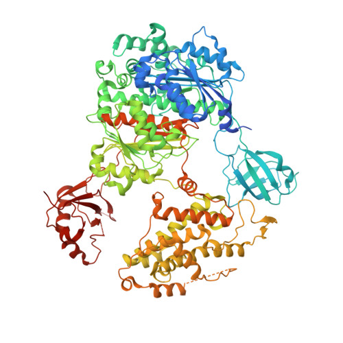
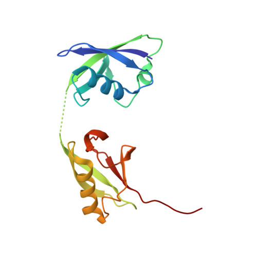
Structures of UBA6 explain its dual specificity for ubiquitin and FAT10.

Truongvan, N.Li, S.Misra, M.Kuhn, M.Schindelin, H.

(2022) Nat Commun 13: 4789-4789

  • DOI: https://doi.org/10.1038/s41467-022-32040-6
  • Primary Citation of Related Structures:  
    7PVN, 7PYV, 7ZH9

  • PubMed Abstract: 

    The covalent modification of target proteins with ubiquitin or ubiquitin-like modifiers is initiated by E1 activating enzymes, which typically transfer a single modifier onto cognate conjugating enzymes. UBA6 is an unusual E1 since it activates two highly distinct modifiers, ubiquitin and FAT10. Here, we report crystal structures of UBA6 in complex with either ATP or FAT10. In the UBA6-FAT10 complex, the C-terminal domain of FAT10 binds to where ubiquitin resides in the UBA1-ubiquitin complex, however, a switch element ensures the alternate recruitment of either modifier. Simultaneously, the N-terminal domain of FAT10 interacts with the 3-helix bundle of UBA6. Site-directed mutagenesis identifies residues permitting the selective activation of either ubiquitin or FAT10. These results pave the way for studies investigating the activation of either modifier by UBA6 in physiological and pathophysiological settings.


  • Organizational Affiliation

    Institute of Structural Biology, Rudolf Virchow Center for Integrative and Translational Bioimaging, University of Würzburg, Josef-Schneider-Straße 2, 97080, Würzburg, Germany.


Macromolecules
Find similar proteins by:  (by identity cutoff)  |  3D Structure
Entity ID: 1
MoleculeChains Sequence LengthOrganismDetailsImage
Ubiquitin-like modifier-activating enzyme 6,Ubiquitin-like modifier-activating enzyme 1,Ubiquitin-like modifier-activating enzyme 6A,
C [auth B]
1,045Homo sapiensMutation(s): 0 
Gene Names: UBA6MOP4UBE1L2UBA1A1S9TUBE1
EC: 6.2.1.45
UniProt & NIH Common Fund Data Resources
Find proteins for P22314 (Homo sapiens)
Explore P22314 
Go to UniProtKB:  P22314
PHAROS:  P22314
GTEx:  ENSG00000130985 
Find proteins for A0AVT1 (Homo sapiens)
Explore A0AVT1 
Go to UniProtKB:  A0AVT1
PHAROS:  A0AVT1
GTEx:  ENSG00000033178 
Entity Groups  
Sequence Clusters30% Identity50% Identity70% Identity90% Identity95% Identity100% Identity
UniProt GroupsA0AVT1P22314
Sequence Annotations
Expand
  • Reference Sequence
Find similar proteins by:  (by identity cutoff)  |  3D Structure
Entity ID: 2
MoleculeChains Sequence LengthOrganismDetailsImage
UBDB [auth C]159Homo sapiensMutation(s): 0 
UniProt & NIH Common Fund Data Resources
Find proteins for O15205 (Homo sapiens)
Explore O15205 
Go to UniProtKB:  O15205
PHAROS:  O15205
GTEx:  ENSG00000213886 
Entity Groups  
Sequence Clusters30% Identity50% Identity70% Identity90% Identity95% Identity100% Identity
UniProt GroupO15205
Sequence Annotations
Expand
  • Reference Sequence
Experimental Data & Validation

Experimental Data

  • Method: X-RAY DIFFRACTION
  • Resolution: 3.27 Å
  • R-Value Free: 0.239 
  • R-Value Work: 0.216 
  • R-Value Observed: 0.217 
  • Space Group: P 1
Unit Cell:
Length ( Å )Angle ( ˚ )
a = 93.458α = 70.519
b = 93.593β = 88.537
c = 109.542γ = 74.593
Software Package:
Software NamePurpose
PHENIXrefinement
XDSdata reduction
XDSdata scaling
PHASERphasing

Structure Validation

View Full Validation Report


Entry History & Funding Information

Deposition Data


Funding OrganizationLocationGrant Number
German Research Foundation (DFG)Germany2243

Revision History  (Full details and data files)

  • Version 1.0: 2022-08-24
    Type: Initial release
  • Version 1.1: 2022-08-31
    Changes: Database references
  • Version 1.2: 2024-01-31
    Changes: Data collection, Refinement description