A recurring packing contact in crystals of InlB pinpoints functional binding sites in the internalin domain and the B repeat.
Geerds, C., Bleymuller, W.M., Meyer, T., Widmann, C., Niemann, H.H.(2022) Acta Crystallogr D Struct Biol 78: 310-320
- PubMed: 35234145 Search on PubMedSearch on PubMed Central
- DOI: https://doi.org/10.1107/S2059798322000432
- Primary Citation Related Structures:
7NMS, 7PV8, 7PV9 - PubMed Abstract:
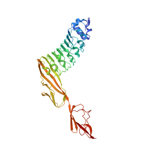
InlB, a bacterial agonist of the human receptor tyrosine kinase MET, consists of an N-terminal internalin domain, a central B repeat and three C-terminal GW domains. In all previous structures of full-length InlB or an InlB construct lacking the GW domains (InlB 392 ), there was no interpretable electron density for the B repeat. Here, three InlB 392 crystal structures in which the B repeat is resolved are described. These are the first structures to reveal the relative orientation of the internalin domain and the B repeat. A wild-type structure and two structures of the T332E variant together contain five crystallographically independent molecules. Surprisingly, the threonine-to-glutamate substitution in the B repeat substantially improved the crystallization propensity and crystal quality of the T332E variant. The internalin domain and B repeat are quite rigid internally, but are flexibly linked to each other. The new structures show that inter-domain flexibility is the most likely cause of the missing electron density for the B repeat in previous InlB structures. A potential binding groove between B-repeat strand β2 and an adjacent loop forms an important crystal contact in all five crystallographically independent chains. This region may represent a hydrophobic `sticky patch' that supports protein-protein interactions. This assumption agrees with the previous finding that all known inactivating point mutations in the B repeat lie within strand β2. The groove formed by strand β2 and the adjacent loop may thus represent a functionally important protein-protein interaction site in the B repeat.
- Department of Chemistry, Bielefeld University, Universitätsstrasse 25, 33615 Bielefeld, Germany.
Organizational Affiliation: