7PTJ

C54S mutant of choline-sulfatase from E. meliloti CECT4857 bound to HEPES


Experimental Data Snapshot

  • Method: X-RAY DIFFRACTION
  • Resolution: 2.10 Å
  • R-Value Free: 0.200 
  • R-Value Work: 0.162 
  • R-Value Observed: 0.164 

wwPDB Validation   3D Report Full Report


Ligand Structure Quality Assessment 


This is version 1.1 of the entry. See complete history


Literature

Structural insights into choline-O-sulfatase reveal the molecular determinants for ligand binding.

Gavira, J.A.Camara-Artigas, A.Neira, J.L.Torres de Pinedo, J.M.Sanchez, P.Ortega, E.Martinez-Rodriguez, S.

(2022) Acta Crystallogr D Struct Biol 78: 669-682

  • DOI: https://doi.org/10.1107/S2059798322003709
  • Primary Citation of Related Structures:  
    6G5Z, 6G60, 7PTH, 7PTJ

  • PubMed Abstract: 

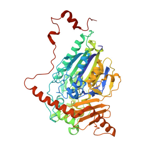
    Choline-O-sulfatase (COSe; EC 3.1.6.6) is a member of the alkaline phosphatase (AP) superfamily, and its natural function is to hydrolyze choline-O-sulfate into choline and sulfate. Despite its natural function, the major interest in this enzyme resides in the landmark catalytic/substrate promiscuity of sulfatases, which has led to attention in the biotechnological field due to their potential in protein engineering. In this work, an in-depth structural analysis of wild-type Sinorhizobium (Ensifer) meliloti COSe (SmeCOSe) and its C54S active-site mutant is reported. The binding mode of this AP superfamily member to both products of the reaction (sulfate and choline) and to a substrate-like compound are shown for the first time. The structures further confirm the importance of the C-terminal extension of the enzyme in becoming part of the active site and participating in enzyme activity through dynamic intra-subunit and inter-subunit hydrogen bonds (Asn146 A -Asp500 B -Asn498 B ). These residues act as the `gatekeeper' responsible for the open/closed conformations of the enzyme, in addition to assisting in ligand binding through the rearrangement of Leu499 (with a movement of approximately 5 Å). Trp129 and His145 clamp the quaternary ammonium moiety of choline and also connect the catalytic cleft to the C-terminus of an adjacent protomer. The structural information reported here contrasts with the proposed role of conformational dynamics in promoting the enzymatic catalytic proficiency of an enzyme.


  • Organizational Affiliation

    Laboratorio de Estudios Cristalográficos, CSIC, Armilla, 18100 Granada, Spain.


Macromolecules
Find similar proteins by:  (by identity cutoff)  |  3D Structure
Entity ID: 1
MoleculeChains Sequence LengthOrganismDetailsImage
Choline sulfatase
A, B, C, D
520Sinorhizobium melilotiMutation(s): 1 
EC: 3.1.6.6
UniProt
Find proteins for O69787 (Rhizobium meliloti (strain 1021))
Explore O69787 
Go to UniProtKB:  O69787
Entity Groups  
Sequence Clusters30% Identity50% Identity70% Identity90% Identity95% Identity100% Identity
UniProt GroupO69787
Sequence Annotations
Expand
  • Reference Sequence
Small Molecules
Ligands 4 Unique
IDChains Name / Formula / InChI Key2D Diagram3D Interactions
EPE (Subject of Investigation/LOI)
Query on EPE

Download Ideal Coordinates CCD File 
E [auth A],
F [auth A],
NA [auth D],
P [auth B],
Z [auth C]
4-(2-HYDROXYETHYL)-1-PIPERAZINE ETHANESULFONIC ACID
C8 H18 N2 O4 S
JKMHFZQWWAIEOD-UHFFFAOYSA-N
SO4
Query on SO4

Download Ideal Coordinates CCD File 
AA [auth C]
BA [auth C]
CA [auth C]
DA [auth C]
EA [auth C]
AA [auth C],
BA [auth C],
CA [auth C],
DA [auth C],
EA [auth C],
G [auth A],
H [auth A],
I [auth A],
J [auth A],
O [auth B],
OA [auth D],
PA [auth D],
Q [auth B]
SULFATE ION
O4 S
QAOWNCQODCNURD-UHFFFAOYSA-L
GOL
Query on GOL

Download Ideal Coordinates CCD File 
FA [auth C]
GA [auth C]
HA [auth C]
IA [auth C]
JA [auth C]
FA [auth C],
GA [auth C],
HA [auth C],
IA [auth C],
JA [auth C],
K [auth A],
KA [auth C],
L [auth A],
LA [auth C],
M [auth A],
R [auth B],
S [auth B],
T [auth B],
U [auth B],
V [auth B],
W [auth B],
X [auth B]
GLYCEROL
C3 H8 O3
PEDCQBHIVMGVHV-UHFFFAOYSA-N
CA (Subject of Investigation/LOI)
Query on CA

Download Ideal Coordinates CCD File 
MA [auth C],
N [auth A],
QA [auth D],
Y [auth B]
CALCIUM ION
Ca
BHPQYMZQTOCNFJ-UHFFFAOYSA-N
Experimental Data & Validation

Experimental Data

  • Method: X-RAY DIFFRACTION
  • Resolution: 2.10 Å
  • R-Value Free: 0.200 
  • R-Value Work: 0.162 
  • R-Value Observed: 0.164 
  • Space Group: C 1 2 1
Unit Cell:
Length ( Å )Angle ( ˚ )
a = 128.521α = 90
b = 207.007β = 110.29
c = 116.833γ = 90
Software Package:
Software NamePurpose
XDSdata reduction
Aimlessdata scaling
PHENIXrefinement
REFMACrefinement
PHASERphasing

Structure Validation

View Full Validation Report


Ligand Structure Quality Assessment 


Entry History & Funding Information

Deposition Data


Funding OrganizationLocationGrant Number
Spanish Ministry of Science, Innovation, and UniversitiesSpainPID2020-116261GB-I00

Revision History  (Full details and data files)

  • Version 1.0: 2022-08-03
    Type: Initial release
  • Version 1.1: 2024-01-31
    Changes: Data collection, Refinement description