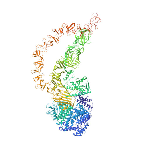
High-resolution structure of native toxin A from Clostridioides difficile.
Aminzadeh, A., Larsen, C.E., Boesen, T., Jorgensen, R.(2022) EMBO Rep 23: e53597-e53597
- PubMed: 34817920 Search on PubMedSearch on PubMed Central
- DOI: https://doi.org/10.15252/embr.202153597
- Primary Citation Related Structures:
7POG - PubMed Abstract:
Clostridioides difficile infections have emerged as the leading cause of healthcare-associated infectious diarrhea. Disease symptoms are mainly caused by the virulence factors, TcdA and TcdB, which are large homologous multidomain proteins. Here, we report a 2.8 Å resolution cryo-EM structure of native TcdA, unveiling its conformation at neutral pH. The structure uncovers the dynamic movement of the CROPs domain which is induced in response to environmental acidification. Furthermore, the structure reveals detailed information about the interaction area between the CROPs domain and the tip of the delivery and receptor-binding domain, which likely serves to shield the C-terminal part of the hydrophobic pore-forming region from solvent exposure. Similarly, extensive interactions between the globular subdomain and the N-terminal part of the pore-forming region suggest that the globular subdomain shields the upper part of the pore-forming region from exposure to the surrounding solvent. Hence, the TcdA structure provides insights into the mechanism of preventing premature unfolding of the pore-forming region at neutral pH, as well as the pH-induced inter-domain dynamics.
- Department of Bacteria, Parasites and Fungi, Statens Serum Institut, Copenhagen, Denmark.
Organizational Affiliation: