The crystal structure of vaccinia virus protein E2 and perspectives on the prediction of novel viral protein folds.
Gao, W.N.D., Gao, C., Deane, J.E., Carpentier, D.C.J., Smith, G.L., Graham, S.C.(2022) J Gen Virol 103
- PubMed: 35020582 Search on PubMedSearch on PubMed Central
- DOI: https://doi.org/10.1099/jgv.0.001716
- Primary Citation Related Structures:
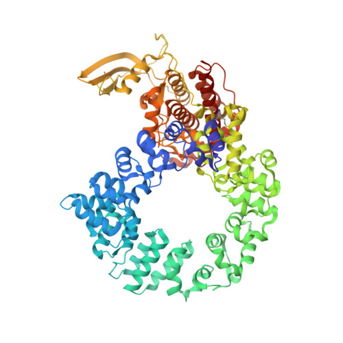
7PHY - PubMed Abstract:
The morphogenesis of vaccinia virus (VACV, family Poxviridae ), the smallpox vaccine, is a complex process involving multiple distinct cellular membranes and resulting in multiple different forms of infectious virion. Efficient release of enveloped virions, which promote systemic spread of infection within hosts, requires the VACV protein E2 but the molecular basis of E2 function remains unclear and E2 lacks sequence homology to any well-characterised family of proteins. We solved the crystal structure of VACV E2 to 2.3 Å resolution, revealing that it comprises two domains with novel folds: an N-terminal annular (ring) domain and a C-terminal globular (head) domain. The C-terminal head domain displays weak structural homology with cellular (pseudo)kinases but lacks conserved surface residues or kinase features, suggesting that it is not enzymatically active, and possesses a large surface basic patch that might interact with phosphoinositide lipid headgroups. Recent deep learning methods have revolutionised our ability to predict the three-dimensional structures of proteins from primary sequence alone. VACV E2 is an exemplar 'difficult' viral protein target for structure prediction, being comprised of multiple novel domains and lacking sequence homologues outside Poxviridae . AlphaFold2 nonetheless succeeds in predicting the structures of the head and ring domains with high and moderate accuracy, respectively, allowing accurate inference of multiple structural properties. The advent of highly accurate virus structure prediction marks a step-change in structural virology and beckons a new era of structurally-informed molecular virology.
- Department of Pathology, University of Cambridge, Tennis Court Road, Cambridge CB2 1QP, UK.
Organizational Affiliation: