Key carboxylate residues for iron transit through the prokaryotic ferritin Syn Ftn.
Bradley, J.M., Fair, J., Hemmings, A.M., Le Brun, N.E.(2021) Microbiology (Reading) 167
- PubMed: 34825885 Search on PubMedSearch on PubMed Central
- DOI: https://doi.org/10.1099/mic.0.001105
- Primary Citation Related Structures:
7PF7, 7PF8, 7PF9, 7PFB, 7PFG, 7PFH, 7PFI, 7PFJ, 7PFK - PubMed Abstract:
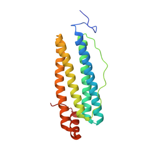
Ferritins are proteins forming 24meric rhombic dodecahedral cages that play a key role in iron storage and detoxification in all cell types. Their function requires the transport of Fe 2+ from the exterior of the protein to buried di-iron catalytic sites, known as ferroxidase centres, where Fe 2+ is oxidized to form Fe 3+ -oxo precursors of the ferritin mineral core. The route of iron transit through animal ferritins is well understood: the Fe 2+ substrate enters the protein via channels at the threefold axes and conserved carboxylates on the inner surface of the protein cage have been shown to contribute to transient binding sites that guide Fe 2+ to the ferroxidase centres. The routes of iron transit through prokaryotic ferritins are less well studied but for some, at least, there is evidence that channels at the twofold axes are the major route for Fe 2+ uptake. Syn Ftn, isolated from the cyanobacterium Synechococcus CC9311, is an atypical prokaryotic ferritin that was recently shown to take up Fe 2+ via its threefold channels. However, the transfer site carboxylate residues conserved in animal ferritins are absent, meaning that the route taken from the site of iron entry into Syn Ftn to the catalytic centre is yet to be defined. Here, we report the use of a combination of site-directed mutagenesis, absorbance-monitored activity assays and protein crystallography to probe the effect of substitution of two residues potentially involved in this pathway. Both Glu141 and Asp65 play a role in guiding the Fe 2+ substrate to the ferroxidase centre. In the absence of Asp65, routes for Fe 2+ to, and Fe 3+ exit from, the ferroxidase centre are affected resulting in inefficient formation of the mineral core. These observations further define the iron transit route in what may be the first characterized example of a new class of ferritins peculiar to cyanobacteria.
- Centre for Molecular and Structural Biochemistry, School of Chemistry, University of East Anglia, Norwich, NR4 7TJ, UK.
Organizational Affiliation: